Product Overview
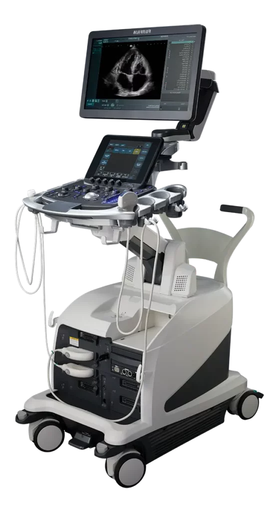
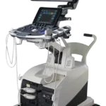
The Fujifilm Lisendo 880 is a portable ultrasound machine that’s designed for cardiology exams. Its small footprint means it can be used in small spaces. Built-in workflows mean exams work quickly, and the resulting images are clean and crisp.
Important features of the Lisendo 880 include the following:
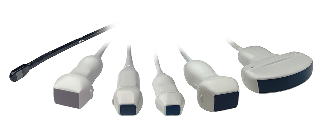
- Transducers designed by cardiologists are optimized for traditional and extensive cardiology exams.
- A protocol assistant increases operator efficiency and ensures appointments move as quickly as possible.
- Several exam types are supported, including vector flow mapping, dual gate Doppler, and more.
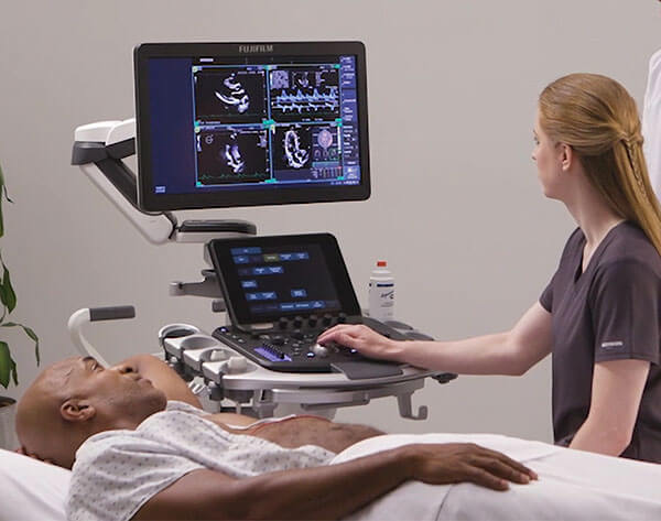
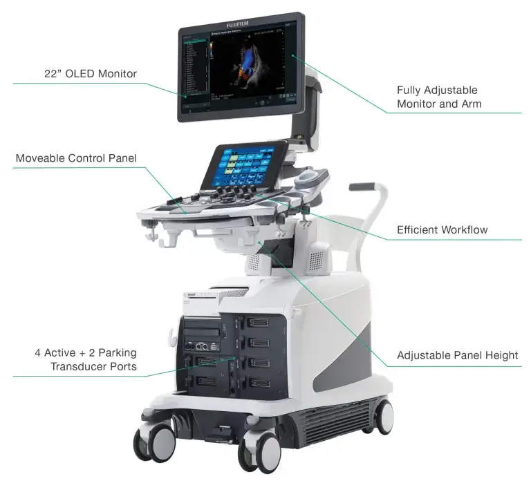
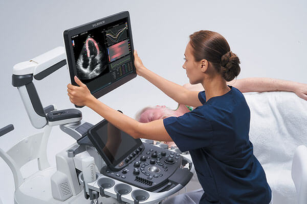
- A fully adjustable 22-inch monitor reduces strain on your operators, even during complex exams.
Core Features of the Lisendo 880
The Lisendo 880 provides crisp cardiology images with improved workflows that reduce inefficiencies. The machine is loaded with ergonomic features that make exams easier on your staff members and patients.
The Lisendo 880 is made specifically for cardiology departments. This machine supports several types of exams, including the following:
- Vector flow mapping to provide an evaluation of the heart’s hemodynamics
- LV eFlow to display blood flow information and improve endocardial border definition
- HemoDynamic Structural Intelligence to measure cardiac anatomy
- Dual Gate Doppler to enable observation of Doppler waveforms from two locations simultaneously
- 3D/4D to provide detailed views of the heart
- 2D tissue tracking to qualify the strain rate of the ventricles
The Lisendo 880 is lightweight and easy to move from place to place. The screen also tilts, swivels, and adjusts for easy viewing during cardiac exams. Your operators will spend less time straining and more time on patient care.
Built-in technology improves workflows and makes exams more efficient. Your technicians can save protocols and imaging conditions, reducing repeat steps. Operators can also customize buttons and personalize their experience based on their preferred history.
The Lisendo 880 allows your team to use pre-set configurations. These steps can save time and make exams much more efficient. Technology also allows for optimized image allocation, reducing operator strain.
Technology
The Lisendo 880 is designed to improve the cardiology exam experience for providers and patients. A crisp monitor that’s easy to adjust and move makes tasks easier for staff. Multiple ports make it easier to switch transducers as needed.
Additional features include the following:
A large monitor makes it easier for your staff members to monitor the exam’s progress and make adjustments as needed. The monitor uses image-enhancing technology to provide crisp details and few shadows.
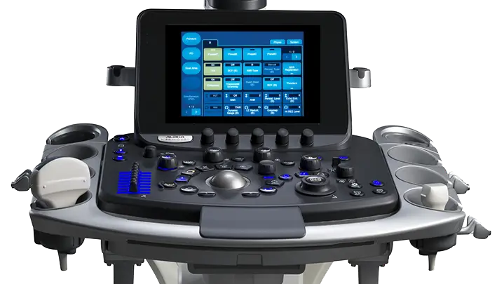
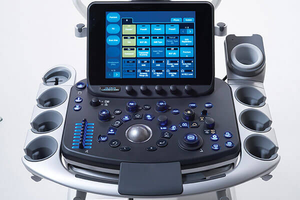
The Lisendo 880 control panel is fully adjustable. Keeping controls within reach makes it easier for your team members to focus on their patients during these difficult exams.
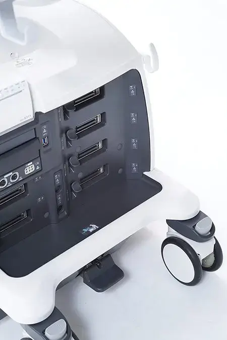
Multiple active transducer ports mean several tools can be connected to the machine and ready for real-time diagnostics. Your team doesn’t need to physically unplug and replug them. Multiple parking ports mean more tools can be connected and organized on the machine.
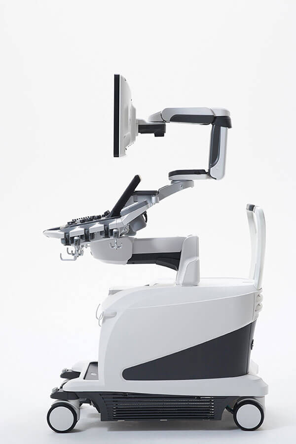
The monitor on the Lisendo 880 can tilt, providing the perfect angle for your technicians, no matter the type of exam your team is delivering. An adjustable arm means your team members can pull the screen forward and pull it back as needed. It features easy mobility and adjustment options.
Technicians can raise or lower the panel’s height very quickly. Tilt and swivel adjustments ensure that your staff members can customize the settings as needed.
Your team members can customize and save specific exam protocols and imaging conditions. With that programming, technicians can reduce the number of manual button operations required during an exam, which can improve efficiencies.
Contact Us Today to Learn More
The Fujifilm Lisendo 880 could be the machine your radiology department needs for efficient and sharp imaging.
At Medical Outfitters, we’ve helped radiology departments make smart decisions about their machines. We can help you understand the benefits of this machine and make a smart purchase choice.
Contact us to get started. We’re ready to talk to you about your needs.
Request a Quote Today
Get in touch with Medical Outfitters today to request your personalized quote and outfit your facility with confidence.

