A neoprobe does something no scanner can. It tells a surgeon, in the middle of an operation and in real time, exactly which piece of tissue is the one they came for. Not an image of roughly where it is, but the tissue itself, identified by the radiation coming out of it.
It is a small device with an outsized role in cancer surgery, and almost nothing is written about it outside manufacturer literature. This guide covers what a neoprobe is, how gamma detection works, what surgeons use it for, and why it is one of the few pieces of theatre equipment with no maintenance schedule at all.
What a Neoprobe Actually Is
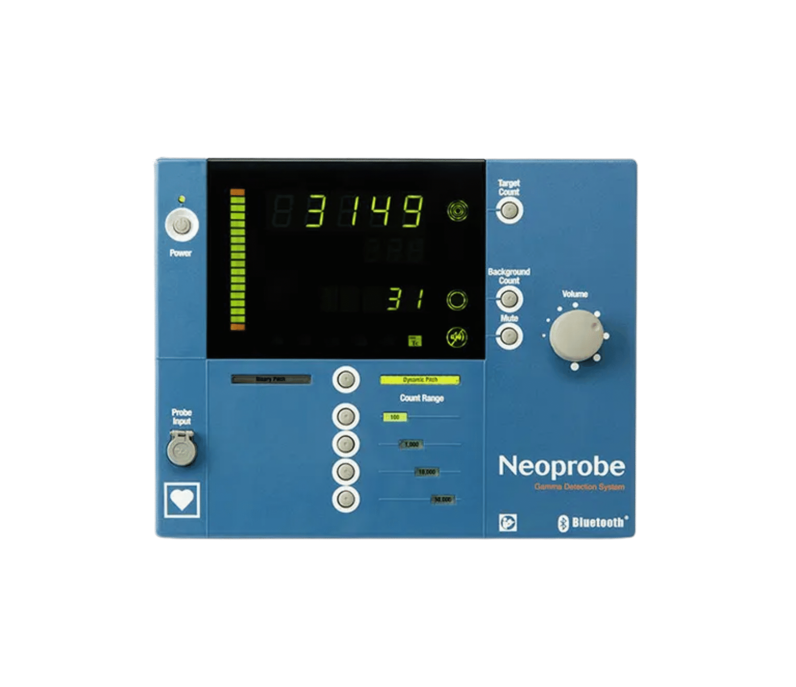
A neoprobe is a handheld gamma detection probe. The Neoprobe Gamma Detection System, made by Mammotome, pairs that probe with a control unit that turns what the probe senses into a number on a screen and a tone in the room. Radioguided surgery is a nuclear medicine procedure, and the Society of Nuclear Medicine and Molecular Imaging is the professional body for the field. Theatre staff usually call the console the Neoprobe machine and the handpiece the Neoprobe probe. Strictly the machine is the control unit and the probe is the part that goes into the wound, and the two are bought and serviced separately.
It is worth being precise about the name. Neoprobe is a brand, not a generic device class; the general term is a gamma detection system, or a gamma probe. The Neoprobe has been the reference product in the category for more than twenty years and is now in its fifth generation, which is why the brand name gets used the way people say Hoover for a vacuum cleaner.
What it is not is an imaging device. It produces no picture. It reports a count rate (how much radiation is reaching the probe tip right now), and it makes a sound whose pitch rises as that number climbs. A surgeon works it by ear, sweeping the probe and listening, rather than by looking at a screen.
How Gamma Detection Works
The technique is called radio-guided surgery, and the sequence is the same whichever procedure it is used for.
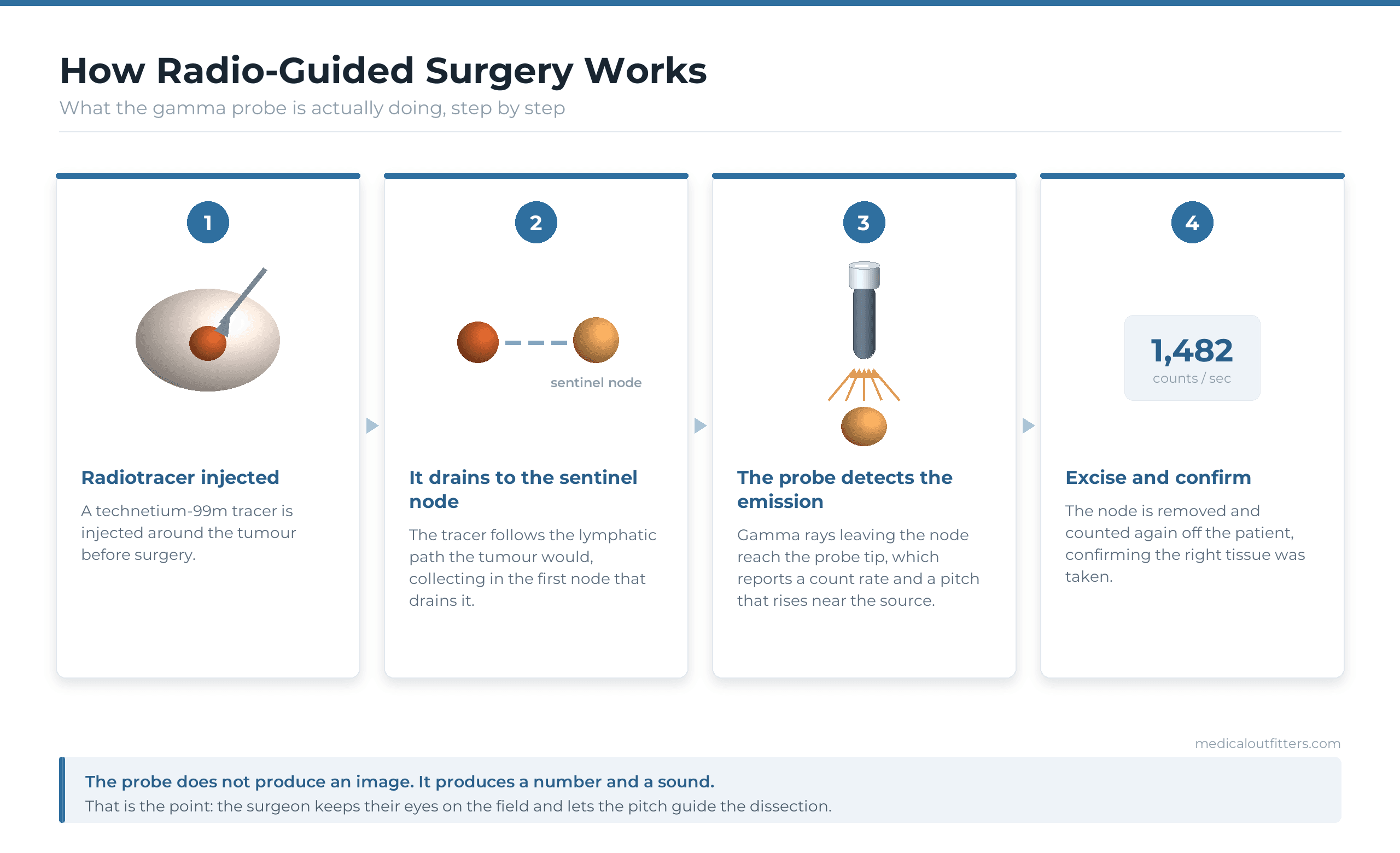
A radiotracer, most commonly one based on technetium-99m, is injected near the tumour before the operation. The tracer travels the lymphatic route the tumour would take, and accumulates in the first node that drains the area. That node is the sentinel node, and it is the one that matters: if cancer has begun to spread, this is where it arrives first.
Why the sentinel node changes the operation
Before this technique existed, finding out whether cancer had spread to the lymph nodes meant removing a large number of them and examining the lot. That carries real long-term consequences for patients, lymphoedema chief among them. Radio-guided surgery lets the surgeon take the one or two nodes that actually answer the question. If those are clear, the rest can usually stay where they are.
How the surgeon knows they have it
The probe is passed over the skin to find the hotspot, then used again inside the wound to guide the dissection. Once the node is out, it is counted again away from the patient to confirm the right tissue was removed, and the surgical bed is re-checked for anything still emitting. A widely used convention is to also take any node counting above roughly ten per cent of the hottest one.
What Surgeons Use a Neoprobe For
Sentinel node biopsy in breast cancer is the largest single use, and it is often done alongside a blue dye so the surgeon has both a visual and an acoustic cue. Melanoma is the other major application, where drainage patterns are far less predictable and a probe is the only practical way to find the right node.
Beyond those two, gamma detection is used in parathyroid surgery to confirm an overactive gland before it is removed, and in radioactive seed localisation, where a tiny iodine-125 seed is placed in a breast lesion too small to feel so the surgeon can find it during excision. It also appears in gynaecological, head and neck, and colorectal work wherever nodal staging is needed.
The Control Unit and the Probes
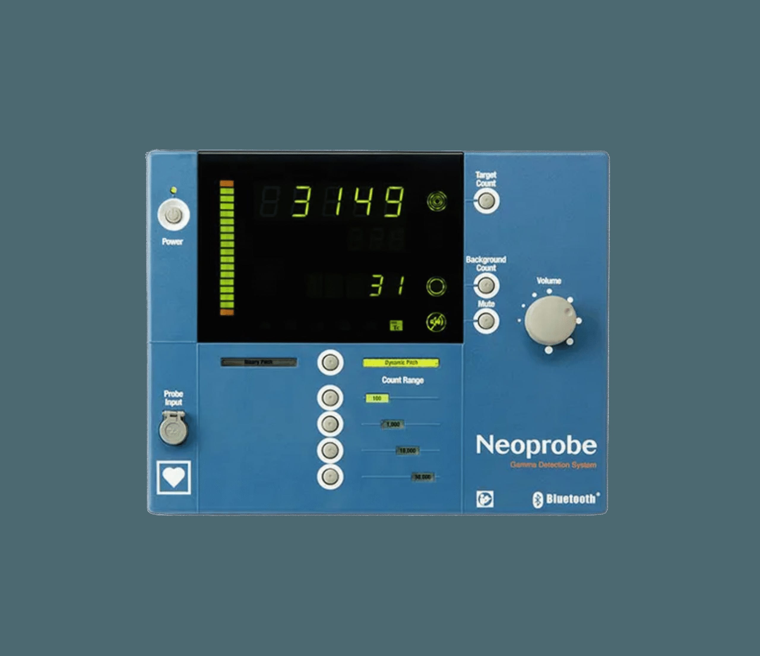
The current Neoprobe system is wireless. The probes connect to the control unit over Bluetooth, which removes the cable that used to tether the surgeon to a console and dictate where they could stand. The console itself detects five isotopes and needs no calibration.
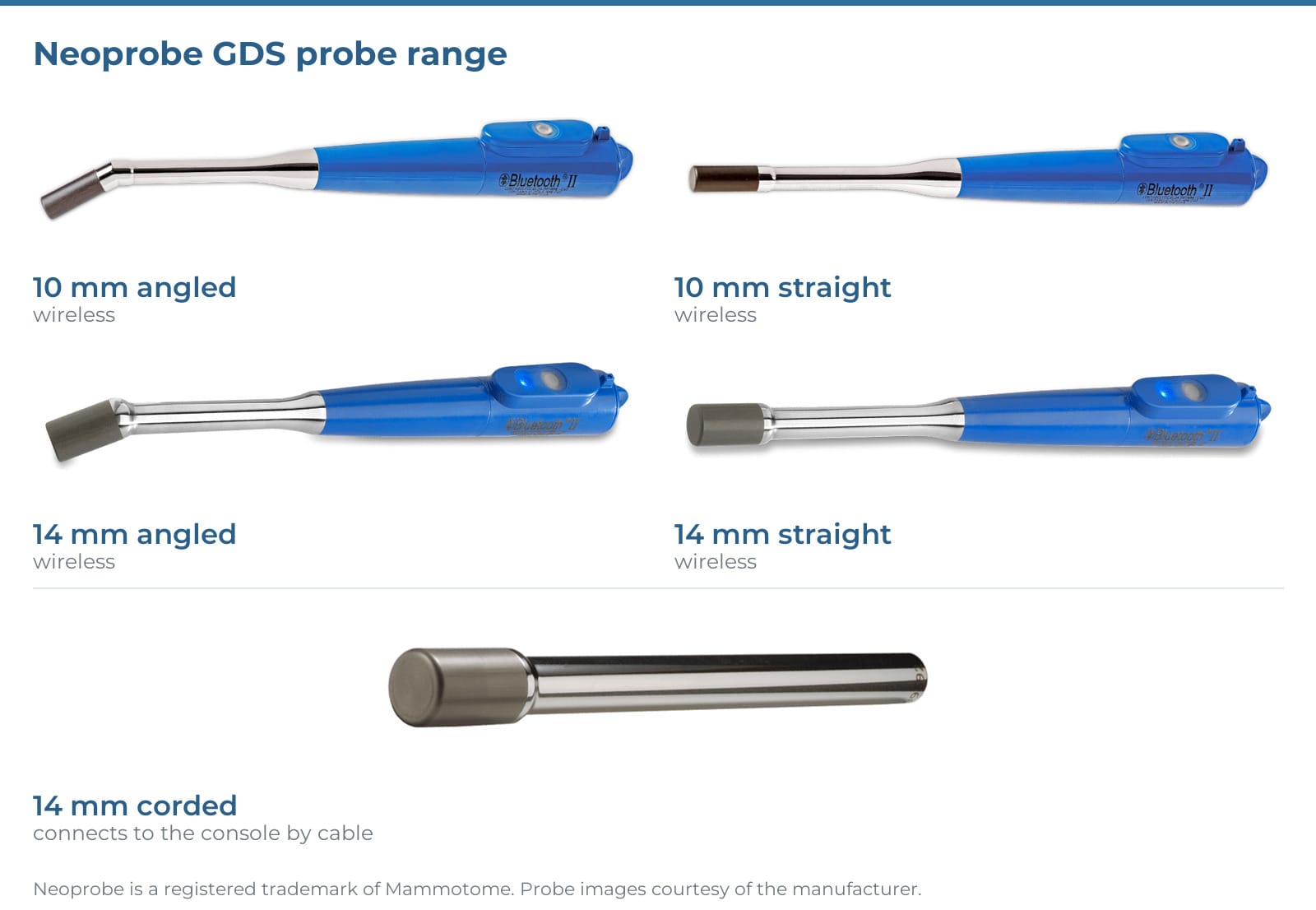
Five probes cover the work, in two families. The 14 mm probes, straight and angled, have the largest head and the highest sensitivity (Mammotome puts them at 50 per cent more sensitive than the corded NPR14), and they can be run with or without an external collimator to narrow the field of view when a hotspot sits close to the injection site. The 10 mm probes, also straight and angled, have a head diameter 28.6 per cent smaller than the 14 mm and are internally collimated for better spatial resolution, which is what you want when incision size matters. A 14 mm corded probe completes the set; it draws power through its cable and works with consoles with or without wireless.
Drag to turn the probe over. Scroll to zoom. Built from Mammotome’s own product photograph, with colours sampled from it.
One feature is easy to overlook and matters more than it sounds: a one-touch remote count that the surgeon can trigger from inside the sterile field. Without it, taking a reading means asking someone else to press a button and waiting.
Dual-Isotope Detection and Why It Matters
The system detects five isotopes (technetium-99m, iodine-125, cobalt-57, indium-111 and iodine-131) plus an open setting, and it can detect two of them at the same time: technetium-99m and iodine-125, with the counts reported separately.
That combination exists for a specific problem. In a breast case using seed localisation, the patient may carry an iodine-125 seed marking the lesion and a technetium-99m tracer mapping the sentinel node, in the same breast, at the same time. A single-isotope probe cannot tell the two signals apart, so the surgeon is left guessing whether a hotspot is the seed or the node. Filtering them in real time removes the ambiguity, and it is the reason a lesion and its sentinel node can be dealt with in one operation.
Why There Is No Maintenance Schedule
This is the part that surprises people who look after imaging equipment for a living. The Neoprobe requires no calibration and has no preventive maintenance interval. It starts instantly and is ready when the theatre needs it.
That is a deliberate design decision rather than a gap in the documentation. Theatre equipment that needs warming up, calibrating or scheduling around does not get used at the moment it is needed. Sealing the detector and removing the calibration requirement is what makes the device dependable in an operating room.
The trade-off is on the other side of the ledger, and it is worth being straight about it: very little of a gamma probe is field repairable. There is no PM schedule to skip, but there is also not much to fix on site when a probe is damaged. Handling is therefore the whole game. Probes are dropped, cables are pulled, and connectors are stressed. Those are the same three failure points that account for most damage to any handheld surgical probe.
- No calibration Nothing to schedule, nothing to drift out of tolerance
- No preventive maintenance interval No PM visit to book or budget for
- Instant startup Ready at the moment the procedure needs it
- Reusable probes Cleaned and reprocessed to the manufacturer’s instructions
- Limited field repair Handling and storage matter more than servicing does
Frequently Asked Questions
What is a neoprobe?
A neoprobe is a handheld gamma detection probe used during surgery to locate tissue that is emitting radiation from an injected radiotracer. Neoprobe is a Mammotome brand name; the generic term is a gamma detection system or gamma probe. It produces no image, only a count rate and an audible tone that rises as the probe nears the source.
What is a gamma detection system used for?
Most commonly sentinel lymph node biopsy in breast cancer and melanoma, where it identifies the first node draining a tumour so only that node needs to be removed. It is also used in parathyroid surgery, in radioactive seed localisation of non-palpable breast lesions, and in gynaecological, head and neck and colorectal procedures that require nodal staging.
How does a neoprobe find a sentinel lymph node?
A technetium-99m radiotracer is injected near the tumour and travels the same lymphatic path the tumour would, collecting in the first draining node. The probe is swept over the skin to find the resulting hotspot, then used inside the wound to guide dissection. After removal the node is counted again off the patient to confirm the correct tissue was taken.
What isotopes does the Neoprobe detect?
Five: technetium-99m, iodine-125, cobalt-57, indium-111 and iodine-131, plus an open setting. It can detect technetium-99m and iodine-125 simultaneously, with the counts filtered and reported separately. That matters when a patient has both an iodine-125 localisation seed and a technetium-99m sentinel node tracer in the same breast during a single operation.
What probes are available for the Neoprobe?
Five: 10 mm straight and angled, 14 mm straight and angled, and a 14 mm corded probe. The 14 mm probes have the highest sensitivity and take an optional external collimator; the 10 mm probes have a head 28.6 per cent smaller and are internally collimated, for procedures where incision size matters.
Does a neoprobe need calibration or preventive maintenance?
No. The Neoprobe Gamma Detection System requires no calibration and has no preventive maintenance interval, and it starts instantly. The trade-off is that very little of a gamma probe is field repairable, so careful handling, correct reprocessing and proper storage matter far more than servicing does.
Where to Go From Here
Medical Outfitters is an authorised Mammotome gamma detection equipment supplier. Full specifications for the system, the complete probe lineup and the isotope detection detail are on the Mammotome Neoprobe product page, and the rest of the range sits in our biopsy and surgical oncology equipment category.
Related Equipment