
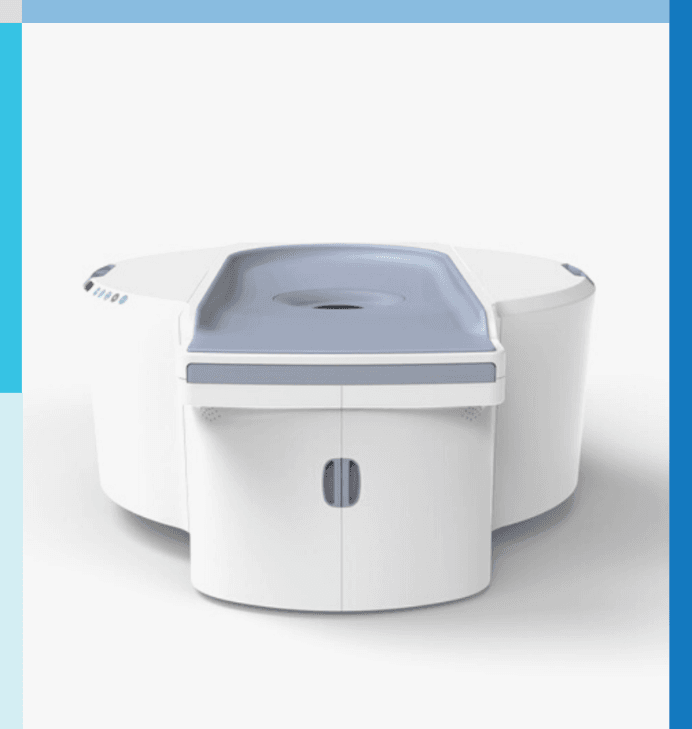
Koning Vera: True 3D Breast CT With No Compression
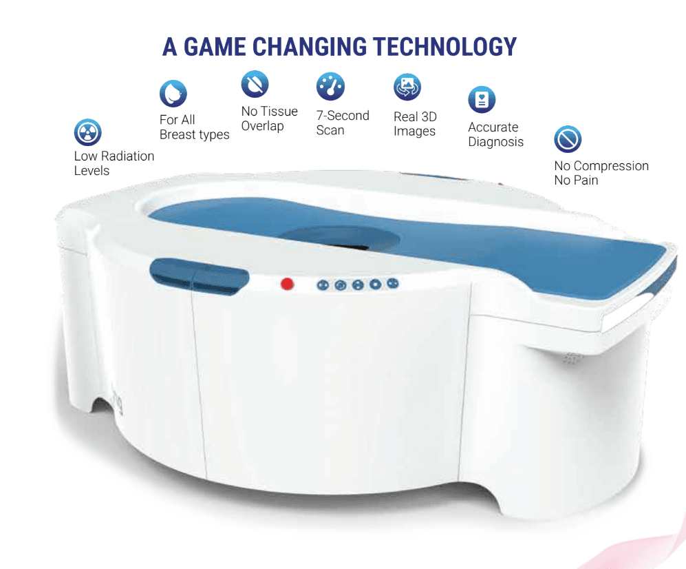
Authorized Koning breast imaging supplier. The Koning Vera, known as KBCT, is the first commercially available dedicated CT scanner designed specifically to image the entire breast, from the chest wall to the nipple. Where traditional two dimensional mammography suffers from structure and tissue overlap that can obscure a lesion, the Koning Vera acquires true 3D isotropic images that remove that overlap and support detection of tumors as small as 2 mm. Koning has developed cone beam CT technology for more than two decades, holds over 80 patents globally, and won the 2016 Frost and Sullivan New Innovation Award in Breast Imaging.
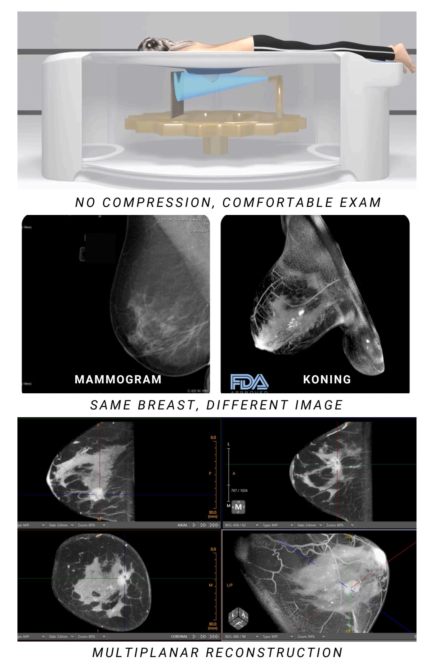
The exam is compression free. The patient lies prone on a dedicated table while a full 3D image is acquired in a short scan, which transforms a procedure that has long been uncomfortable for patients. A self-shielding design and an integrated operator console remove the need for a separate control room, the table elevates up to about 1.55 meters, and an optional biopsy kit allows biopsies directly on the table. Images are DICOM compliant and connect to most RIS and PACS systems for remote viewing.
Need pricing? Get a quote with lead times and financing options within one business day.
True 3D Imaging, Compression Free Comfort, and On-Table Biopsy
Three qualities define the Koning Vera. True 3D isotropic cone beam imaging removes the tissue overlap of mammography, a compression free prone exam transforms patient comfort, and a self-shielding design with an optional biopsy kit fits into existing rooms and workflow. Full technical detail is in the expandable sections below.
Full 3D isotropic imaging: the entire breast is imaged from the chest wall to the nipple
No tissue overlap: true 3D acquisition removes the structure and tissue overlap that can hide a lesion on 2D mammography
Small lesion detection: supports detection of tumors as small as 2 mm
High resolution reconstruction: standard voxel size of 0.273 mm, with a high resolution option of 0.190 and 0.155 mm
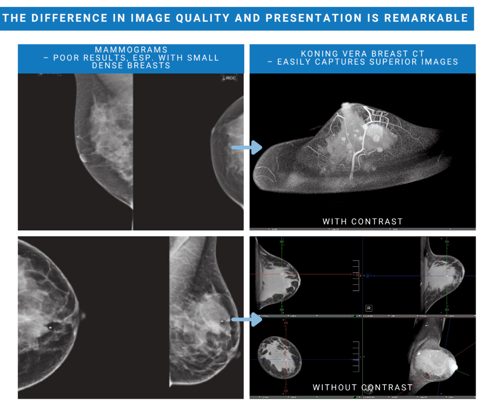
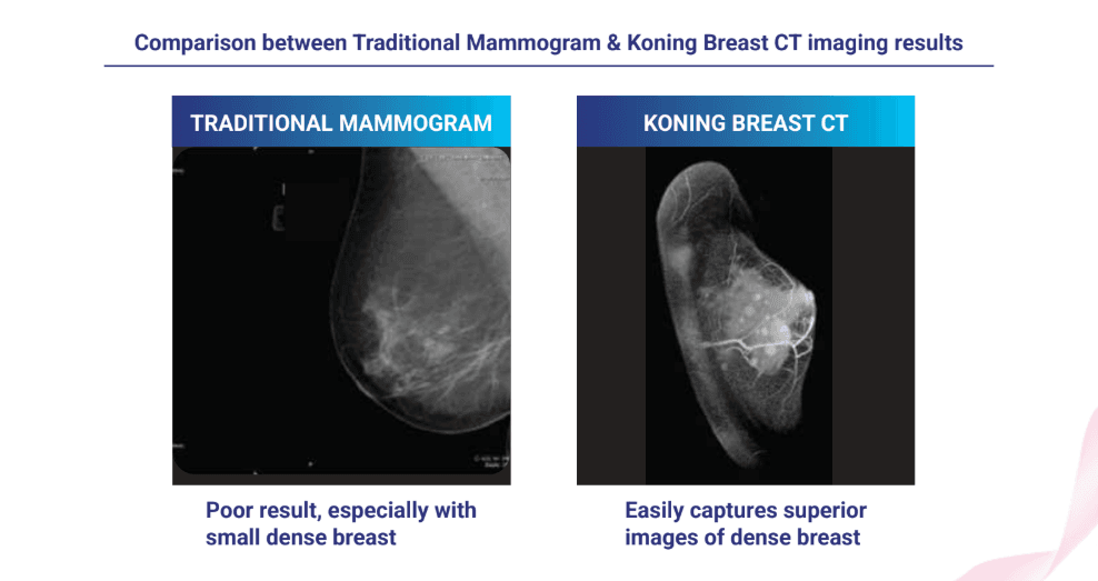
For all breast types: captures superior images of dense breast tissue where mammography can give poor results
No compression and no pain: the exam requires no breast compression, unlike mammography which compresses the breast with about 10 to 20 kg of force
Prone positioning: the patient lies prone on a unique dedicated table for an improved patient experience
Wide patient access: interlocking safety covers on both sides provide access to the patient
On-table biopsy: the table elevates up to about 1.55 meters and, with the optional biopsy kit, supports biopsies directly on the table, which can remove the need for a separate stereotactic biopsy table
Surgical planning: full 3D images help breast surgeons plan procedures based on 3D anatomy
How the Koning Vera compares against traditional mammography:
On a two dimensional mammogram, overlapping breast structures can hide a small cancer and reduce contrast, which is one reason mammography can fall short on early and dense breast cancers. Because the Koning Vera reconstructs the breast as a true 3D isotropic volume, structures are separated in space rather than projected on top of one another, which removes overlap and supports detection of tumors as small as 2 mm. The same volumetric data set gives breast surgeons a 3D view of the anatomy for surgical planning.
Full System Specifications
Complete reference data for procurement, site planning, and clinical review. Imaging directors, radiologists, and facilities teams rely on this specification set when they evaluate dedicated breast CT systems. Expand each category for full detail.
| Image Type | True 3D isotropic cone beam breast CT |
| Coverage | Entire breast, from the chest wall to the nipple |
| Scan Time | 10 seconds, 300 projections per scan |
| Standard Reconstruction | Voxel size of 0.273 mm |
| High Resolution Reconstruction | Voxel size of 0.190 and 0.155 mm |
| Lesion Detection | Supports detection of tumors as small as 2 mm |
| Air Kerma | 25 mGy, with a tolerance of 20 percent |
| Half Value Layer | Greater than 0.49 mm Al at 49 kVp, which is the FDA specification |
| Half Value Layer, typical | 1.5 mm Al, with a tolerance of 10 percent, at 49 kVp |
| Relative Dose | Equivalent radiation dose to mammography |
| Patient Position | Prone on a dedicated exam table |
| Patient Table Load | 200 kg maximum |
| Patient Table Height | 1.0 m at the minimum position to 1.55 m at the maximum position, with a tolerance of 10 percent |
| Patient Access | Wide interlocking safety covers on both sides |
| Biopsy | Optional biopsy kit for biopsies directly on the table |
| Shielding | Self-shielding design with integrated operator console, no separate control room required |
| Room Fit | Sized to fit into standard stereotactic rooms |
| Minimum Room Size | 5.5 m by 6.0 m, recommended |
| Operating Temperature | 20 °C to 24 °C |
| Humidity | 30 percent to 60 percent relative humidity, non-condensing |
| Input Voltage and Current | 480 V three phase at 60 A, or 208 V three phase at 120 A, plus ground |
| Maximum Voltage | 49 kVp |
| Maximum Current | 200 mA |
| Power Output | 9.8 kW |
Clinical Applications
The Koning Vera supports breast imaging across screening, diagnosis, intervention, and surgical planning, in breast centers, radiology departments, and hospitals.
True 3D isotropic imaging captures superior images of dense breast tissue, where two dimensional mammography often gives poor results.
By removing tissue overlap, the Koning Vera supports detection of tumors as small as 2 mm for earlier breast cancer detection.
A prone, compression free exam offers a more comfortable alternative for patients who find mammography painful.
True 3D images help characterize a suspicious lesion without the structure overlap of a flat mammogram.
With the optional biopsy kit, biopsies are performed directly on the elevated table, which can remove the need for a separate stereotactic table.
Full 3D volumetric images give breast surgeons a clear view of the anatomy for planning procedures.
From Prone Positioning to Surgical Planning
The Koning Vera is designed to fit a full breast imaging pathway on a single device, from a comfortable compression free scan through DICOM reading, on-table biopsy, and surgical planning. The stages below outline the workflow.
The patient lies prone on the Koning Vera dedicated exam table with the breast positioned for imaging, and no compression is applied. Wide interlocking safety covers on both sides give access to the patient. The prone, compression free setup is designed to make the exam comfortable for what has long been a difficult procedure.
The system acquires a full 3D image of the breast with 300 projections per scan and a scan time of 10 seconds. Data reconstructs into true 3D isotropic images at a standard voxel size of 0.273 mm, or at a high resolution voxel size of 0.190 and 0.155 mm, which supports detection of tumors as small as 2 mm.
The images generated are DICOM compliant and plug directly into most RIS and PACS systems for remote viewing, so radiologists can read studies on their existing reading workstations and breast imaging monitors.
The table elevates up to about 1.55 meters, and with the optional biopsy kit a biopsy can be performed directly on the table. This can eliminate the need for a separate stereotactic biopsy table and keeps the patient on a single device.
Because the study is a true 3D volume, breast surgeons can review the anatomy in three dimensions and plan procedures based on the spatial relationships of the tissue, rather than on flat 2D projections.
Connectivity, Quality, and Support
The Koning Vera produces DICOM compliant images that connect to RIS and PACS, is built on more than two decades of cone beam CT development with over 80 patents, and ships with training, lifetime service and parts warranties, and flexible financing. Medical Outfitters is an ISO 13485:2016 certified supplier, so every system ships through a documented and audited quality management system. Full detail is in the expandable sections below.
The images generated by the Koning Vera are DICOM compliant and plug directly into most RIS and PACS systems for remote viewing, so studies integrate with the existing reading and archiving environment.
Koning has been developing cone beam CT technology for more than two decades and holds over 80 patents globally. The technology was recognized with the 2016 Frost and Sullivan New Innovation Award in Breast Imaging.
Room fit: the size, dimensions, and self-shielding allow the system to fit into standard stereotactic rooms, which avoids costly construction.
Training: technician training is provided with each install.
Warranty: service and parts warranties are offered for the life of the product, and a range of financing options is available.
Medical Outfitters Inc. is an independently ISO 13485:2016 certified supplier of medical imaging equipment. Every Koning Vera order is fulfilled through a documented and audited quality management system.
Site Planning & Installation Review
Share your available room and whether you have an existing stereotactic suite. Our team confirms whether the self-shielding Koning Vera fits your space, plans the electrical and HVAC requirements, and arranges technician training before your order ships.
Breast Center Procurement & Financing
Volume pricing, technical documentation, and flexible financing from lease buy-back to per-scan revenue sharing. We support radiologists, breast surgeons, and purchasing teams sourcing dedicated breast CT. Contact us for RFP packages.
Frequently Asked Questions
Quick answers to the most common purchase, clinical, and siting questions about the Koning Vera 3D Breast CT. Tap any question to expand.
The Koning Vera, also called KBCT, is a dedicated 3D breast CT system. It is the first commercially available dedicated CT scanner designed specifically to image the entire breast from the chest wall to the nipple. It uses cone beam CT technology to acquire true 3D isotropic images with the patient lying prone and with no breast compression.
Traditional mammography is a two dimensional technique, so overlapping structures and tissue can obscure a lesion, and the exam requires the breast to be compressed with about 10 to 20 kg of force. The Koning Vera acquires true 3D isotropic images, which removes that structure and tissue overlap and supports detection of tumors as small as 2 mm, and it does so without any compression.
No. The Koning Vera is a compression free exam. The patient lies prone on a dedicated table while the breast is imaged, which is designed to be far more comfortable than compression based mammography, and it works for all breast types, including small and dense breasts.
The system acquires a full 3D image in a short scan. The technical specification lists a scan time of 10 seconds with 300 projections per scan. Images reconstruct at a standard voxel size of 0.273 mm, with a high resolution option of 0.190 and 0.155 mm, which supports detection of tumors as small as 2 mm.
Koning states that the radiation dose is equivalent to mammography. The specification lists an air kerma of about 25 mGy with a tolerance of 20 percent, and a half value layer greater than 0.49 mm of aluminum at 49 kVp, which meets the FDA specification value.
Yes. The table elevates up to about 1.55 meters and, with the optional biopsy kit, biopsies can be performed directly on the table. This can eliminate the need for a separate stereotactic biopsy table. The full 3D images are also useful to breast surgeons for planning surgery based on 3D anatomy.
The self-shielding design and integrated operator console remove the need for a separate control room, and the system is sized to fit into standard stereotactic rooms, which avoids costly construction. The minimum recommended room size is 5.5 m by 6.0 m. Electrical input is 480 V three phase at 60 A, or 208 V three phase at 120 A, plus ground, with a maximum power output of 9.8 kW.
The Koning Vera is available through Medical Outfitters Inc., an ISO 13485:2016 certified supplier based in Miami, FL. We provide site planning, technician training support, and nationwide installation. Call (305) 885-4045 or request a quote online.
Compatible Equipment
Breast imaging equipment commonly deployed alongside the Koning Vera, from a digital mammography system and a vacuum-assisted biopsy platform to the high resolution mammography monitor that radiologists read the studies on.
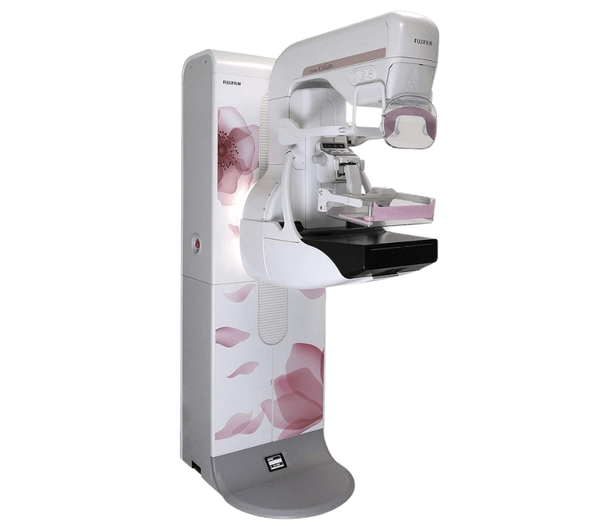
Fujifilm ASPIRE Cristalle
Digital mammography · tomosynthesis · companion screening modality
View Product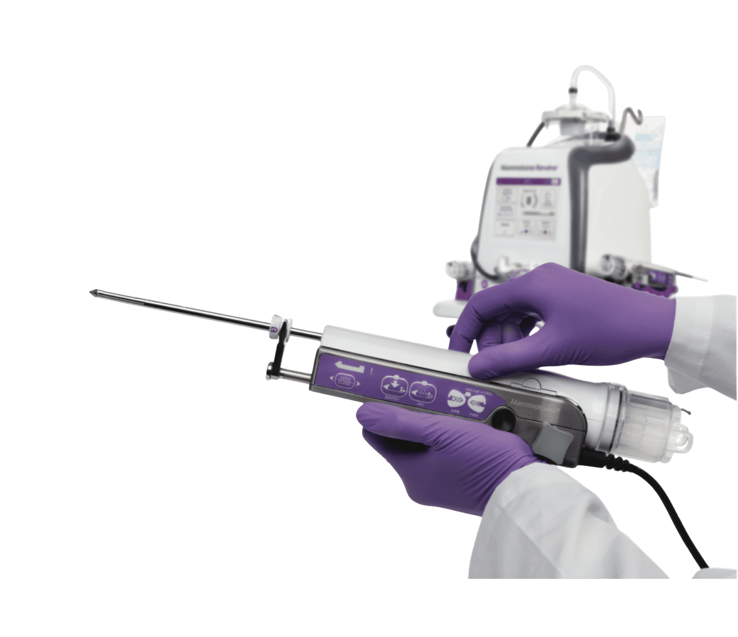
Mammotome Revolve Biopsy System
Dual vacuum-assisted breast biopsy · tissue acquisition · image guided
View Product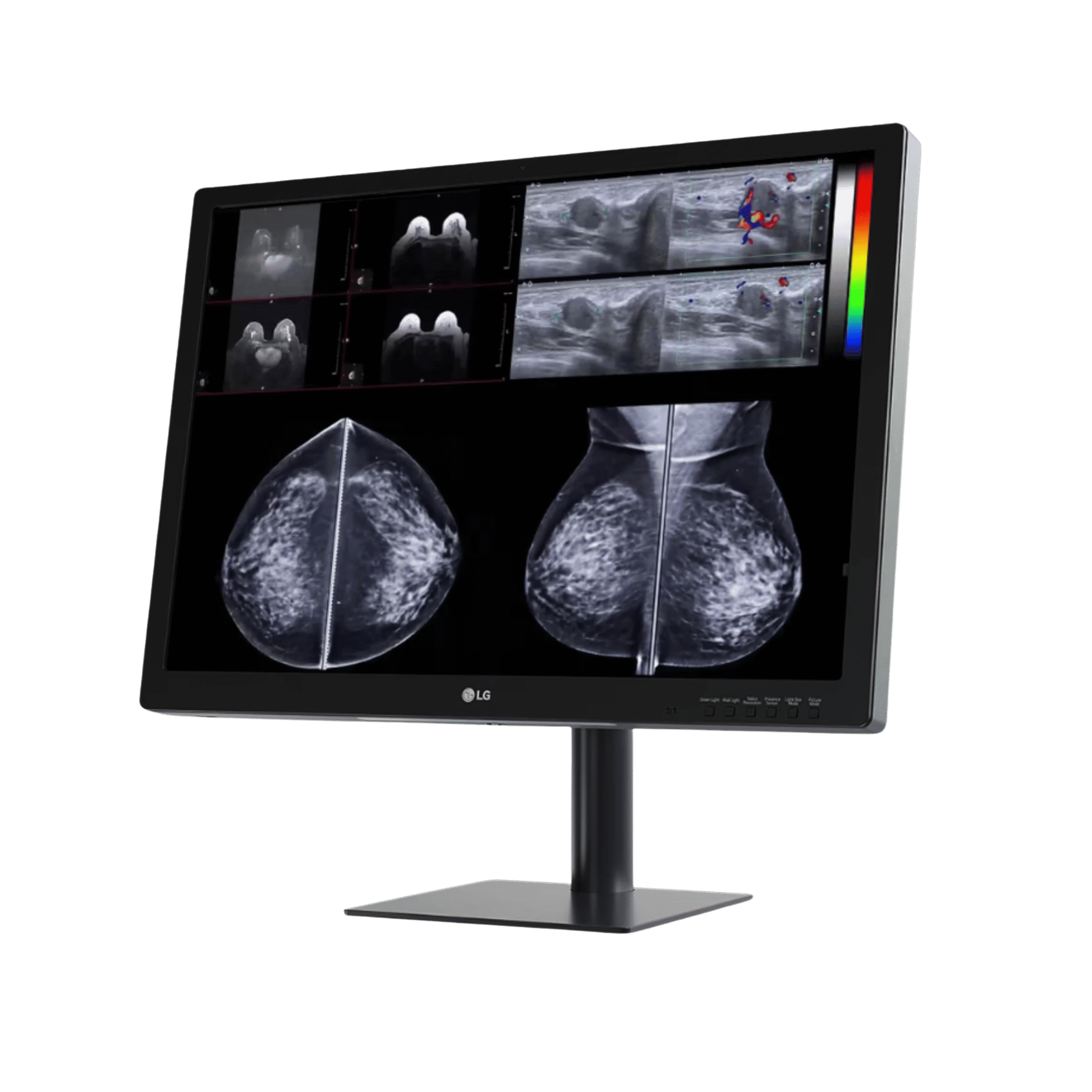
LG 31HN713D Mammography Monitor
31" 12MP IPS · reads DICOM breast studies · diagnostic review
View ProductRequest a Quote Today
Get in touch with Medical Outfitters today to request your personalized quote and outfit your facility with confidence.


