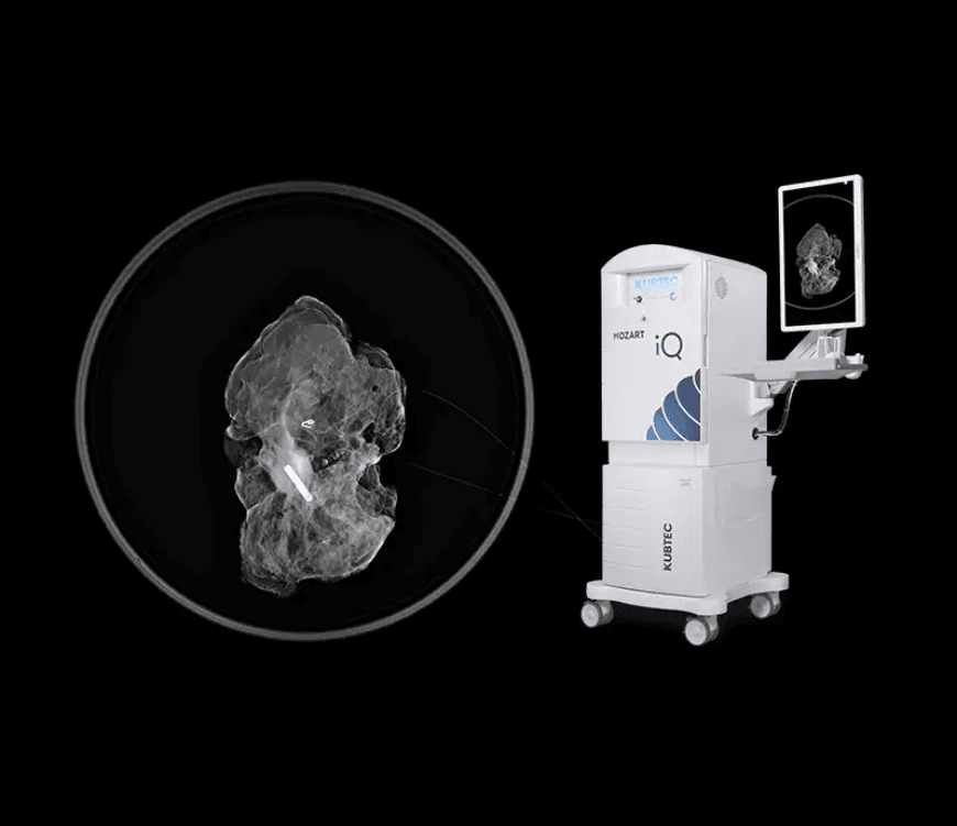
Kubtec MOZART Specimen Tomosynthesis System: 3D Margin Assessment in the Operating Room
Authorized Kubtec specimen tomosynthesis equipment supplier. The MOZART System represents the pinnacle of breast cancer margin assessment, using intraoperative 3D tomosynthesis to identify surgical margins in three axes. As a major advancement from KUBTEC, it empowers surgeons to close with confidence by visualizing significantly more information than traditional 2D specimen imaging, directly in the operating room.
By using the same gold standard 3D X-ray technology found in diagnostic mammography, the MOZART provides an exceptionally accurate method for detecting positive margins during breast conserving surgery and segmental mastectomies. Specimen tomosynthesis reconstructs the specimen in 1 millimeter digital slices, each independent of the others and unobscured by dense tissue above or below, so the surgeon can analyze the location and extent of a lesion and the involvement of the peripheral, anterior, and posterior margins. An HD optical camera with the Image Blender, AutoMagnification, voice control, and an automatic specimen alert round out a system built for the surgical suite.
Need pricing? Get a quote with lead times and volume discounts within one business day.
Intraoperative 3D Tomosynthesis, 1 Millimeter Slices, and OR Tools
The MOZART brings 3D specimen tomosynthesis into the surgical suite. Independent 1 millimeter slices, true three axis margin analysis, and operating room focused tools give the surgeon confidence to close at the index operation. Full technical detail is in the expandable sections below.
Intraoperative 3D Tomosynthesis: uses 3D digital breast tomosynthesis to show surgical margins with high accuracy in the operating room, the next gold standard for specimen mammography
1 Millimeter Digital Slices: reconstructs the specimen into 1 millimeter slices, each anatomically with its own margin and viewable independently of the others
Unobscured View: each slice is unobscured by dense tissue above or below, which removes the projection errors of a single planar 2D view
Margin Analysis in Three Axes: analyze the location and extent of a lesion and the involvement of the peripheral, anterior, and posterior margins
Direct Capture Detail: high contrast direct capture imaging identifies microcalcifications, marker seeds, and biopsy clips and their true proximity to each margin
Targeted Shave at the Index Operation: the surgeon can excise additional margin during the first operation, reducing the need for a second procedure
HD Optical Camera and Image Blender: the Image Blender dynamically overlays the X-ray and optical images of the specimen so the team can locate lesions and markers directly on the physical specimen, a tool designed for the operating room
AutoMagnification: automatic magnification brings small structures into clear view without manual adjustment
Voice Control: speech recognition supports a fast, hands-free review at the surgical point of care
Automatic Specimen Alert: a safety feature that notifies the team if a specimen is left inside the system
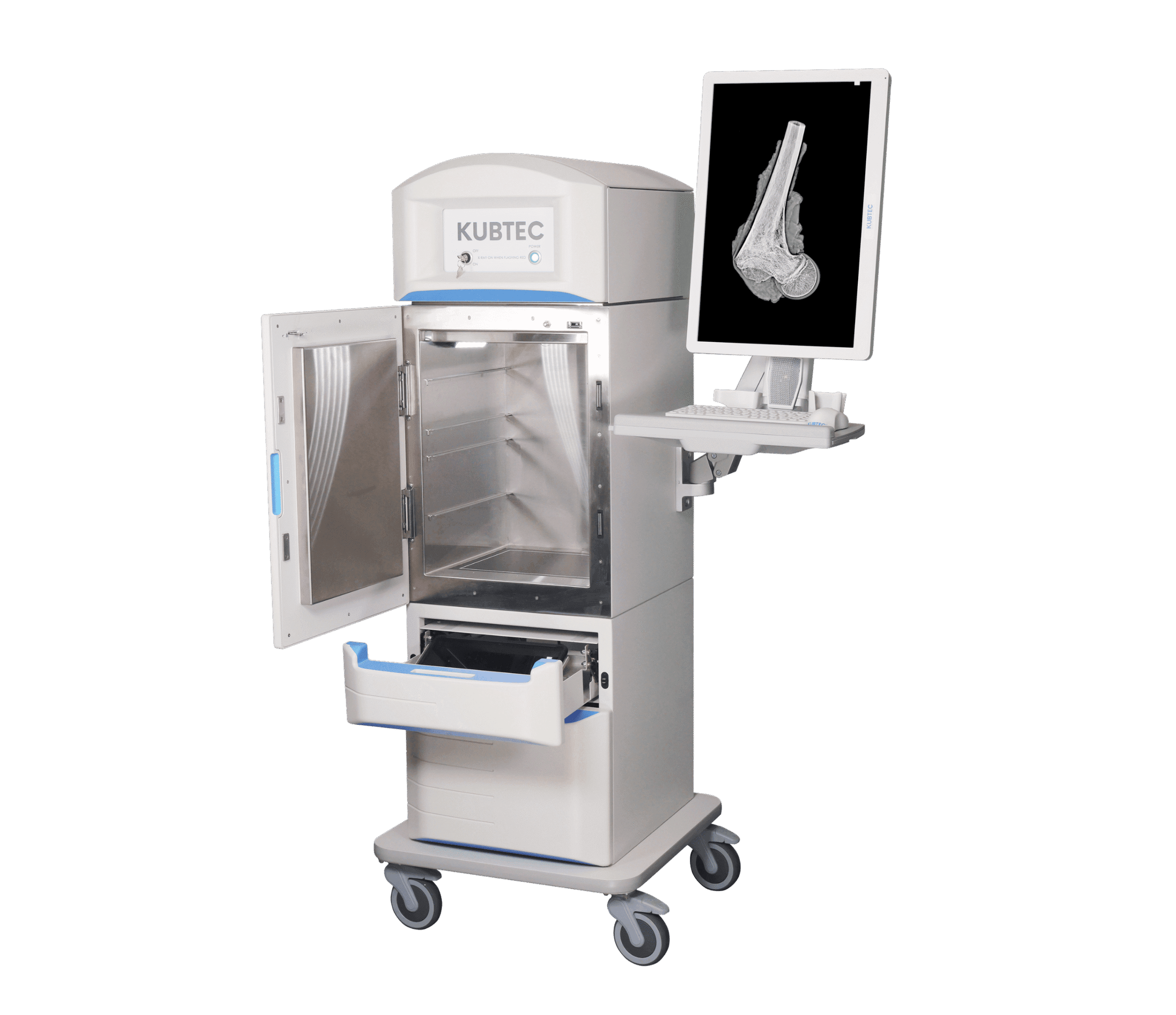
MOZART and MOZART Supra: available in MOZART and larger MOZART Supra configurations to match specimen size and operating room needs
Integrated Probe Option: an integrated gamma probe option supports seamless localization workflows alongside specimen imaging
How the MOZART specimen tomosynthesis compares against a traditional 2D specimen X-ray:
The advantages of the MOZART are backed by leading clinical institutions. A UT Southwestern study found that intraoperative 3D tomosynthesis is independently associated with a re-excision rate reduction of more than 50 percent compared with 2D imaging. A Rush University study reported an average of 7.6 minutes saved per surgery and an estimated operating room cost savings of 284.62 dollars per case for wire localized segmental mastectomies with sentinel node biopsy. MD Anderson research found that 3D tomosynthesis is less likely to recommend excising additional healthy tissue unnecessarily, which helps preserve cosmesis while confirming clear margins in the operating room.
Full Technical Specifications
Complete reference data for capital procurement, RFP submissions, and surgical and biomedical review. Breast surgeons, operating room directors, and biomedical engineers rely on this specification set when they evaluate an intraoperative specimen tomosynthesis system. Expand each category for full detail.
| Imaging Mode | 3D digital breast tomosynthesis specimen imaging |
| Slice Reconstruction | 1 millimeter independent digital slices |
| Active Imaging Area | 6 by 8 inch active imaging area for mastectomies and large specimens |
| Detector | Direct capture detector for high contrast specimen images |
| Acquisition | Rapid acquisition of high resolution cross sectioned images of the intact specimen |
| Visualized Structures | Microcalcifications, marker seeds, biopsy clips, and lymph nodes |
| Margin Axes | Surgical margins identified in three axes |
| Slice Review | Each slice has its own margin and is viewed independently |
| Margins Assessed | Peripheral, anterior, and posterior margins |
| Tissue Clarity | View of each slice unobscured by dense tissue above or below |
| Decision Support | Supports targeted shave decisions at the index operation |
| HD Optical Camera | Integrated high definition optical camera of the physical specimen |
| Image Blender | Dynamically overlays X-ray and optical images of the surgical specimen |
| AutoMagnification | Automatic magnification of small structures |
| Voice Control | Speech recognition for hands-free operation in the operating room |
| Automatic Specimen Alert | Notifies the team if a specimen is left inside the system |
| Cabinet Design | Self contained X-ray cabinet system for safe specimen imaging |
| Connectivity | Images integrate with imaging information systems through standard connectivity |
| Models | MOZART System and larger MOZART Supra System |
| Application | Intraoperative specimen imaging for breast conserving surgery and segmental mastectomy |
| Integrated Probe Option | Integrated gamma probe option for localization workflows |
| Manufacturer | KUB Technologies, Inc. |
Clinical Applications
Deployed in the operating room where intraoperative 3D specimen imaging, true margin analysis, and real time decision making determine whether a breast conserving operation is complete at the index procedure.
3D specimen tomosynthesis shows surgical margins in three axes in the operating room, so the team can confirm clear margins before closing.
Real time review of 1 millimeter slices during lumpectomy helps the surgeon evaluate lesion location and margin involvement at the index operation.
The large active imaging area and tomosynthesis reconstruction suit wire localized segmental mastectomy specimens.
By revealing positive margins during surgery, the MOZART supports a targeted shave that has reduced re-excision rates by more than 50 percent in clinical studies.
Independent slices pinpoint how far microcalcifications, marker seeds, and clips extend from each margin inside the specimen.
The HD optical camera and Image Blender let the team correlate imaging with the physical specimen without leaving the operating room.
From Excised Specimen to Confident Closure
An operating room workflow built to confirm clear margins during surgery. On-site tomosynthesis, three axis margin analysis, and targeted shave decisions move the case from excision to closure without a second procedure. Expanded workflow details are in the expandable sections below.
The excised breast specimen is imaged in the operating room rather than sent to diagnostic radiology, which keeps the surgeon in control of the decision and saves operating room time. Intraoperative imaging on average shortened operating room time by 7.6 minutes compared with sending the specimen out.
The MOZART performs rapid 3D digital breast tomosynthesis and reconstructs the specimen into 1 millimeter slices in roughly a minute. Each slice is independent and unobscured by dense tissue above or below, which reveals the true location of a lesion and any microcalcifications relative to every margin.
The surgeon reviews the peripheral, anterior, and posterior margins slice by slice, while the HD optical camera and the Image Blender overlay the X-ray and optical images on the physical specimen. AutoMagnification and voice control support a fast, hands-free read at the point of care.
If a margin is involved, the surgeon performs a targeted shave at the index operation rather than scheduling a re-excision, and the automatic specimen alert helps ensure no specimen is left inside the system. The result is fewer reoperations, preserved healthy tissue, and better cosmesis for the patient.
Regulatory Compliance and Safety
FDA cleared for specimen imaging, manufactured by KUB Technologies, Inc. Medical Outfitters is an ISO 13485:2016 certified supplier of imaging equipment, so every Kubtec MOZART ships through a documented and audited quality management system. Full compliance documentation is available in the expandable sections below.
FDA Cleared: the Kubtec MOZART specimen tomosynthesis system is FDA cleared for specimen imaging.
ISO 13485: Medical Outfitters Inc. is independently ISO 13485:2016 certified as a supplier, so every system ships through a fully audited quality management system.
Radiation Safety: the MOZART is a self contained X-ray cabinet system designed for safe intraoperative specimen imaging in the surgical suite.
Trademarks: Kubtec, the Kubtec logo, MOZART, Supra, and Image Blender are registered trademarks of KUB Technologies, Inc.
Connectivity: images integrate with imaging information systems through standard connectivity for the surgical and pathology record.
RoHS: Restriction of Hazardous Substances compliant.
REACH: EU REACH chemical regulation compliant.
Operating Room & Workflow Review
Tell us your surgical volume and operating room layout. Our team confirms the right configuration, including the MOZART or MOZART Supra and connectivity, and coordinates installation and support.
Capital Equipment & RFP Support
Pricing, technical documentation, and global logistics for specimen tomosynthesis deployments. We support breast surgery directors, biomedical engineers, and purchasing departments evaluating intraoperative imaging systems. Contact us for RFP packages.
Frequently Asked Questions
Quick answers to the most common purchase and clinical questions about the Kubtec MOZART specimen tomosynthesis system. Tap any question to expand.
The Kubtec MOZART is an intraoperative specimen tomosynthesis system that brings 3D digital breast tomosynthesis into the operating room for surgical margin assessment. It reconstructs the breast specimen in 1 millimeter digital slices, which lets the surgeon evaluate the location and extent of a lesion and the involvement of the peripheral, anterior, and posterior margins in real time during breast conserving surgery.
A traditional 2D specimen X-ray compresses the three dimensional anatomy of the specimen into a single planar view, so all vertical perspective is lost and a positive margin can be missed until final pathology. Specimen tomosynthesis instead creates independent 1 millimeter slices, each with its own margin, so the view of each slice is unobscured by dense tissue above or below and margins are shown as they really are.
By revealing the true location of a lesion relative to every margin during surgery, the MOZART lets the surgeon perform a targeted shave at the index operation rather than scheduling a second procedure. Clinical studies report that intraoperative 3D tomosynthesis reduces re-excision rates by more than 50 percent compared with 2D imaging, with one center reducing its rate from 16 percent to 9 percent.
The MOZART combines an HD optical camera with the Image Blender, which dynamically overlays the X-ray and optical images of the surgical specimen. This lets the team identify the location of lesions and markers directly on the physical specimen, while AutoMagnification and voice control support a fast, hands-free review at the point of care in the operating room.
Specimen tomosynthesis reconstructs the specimen into 1 millimeter slices that can each be viewed independently. Because each slice anatomically has its own margin, the surgeon can pinpoint how far microcalcifications or a lesion extend from a given margin, for example confirming that calcifications reach within 1 millimeter of the anterior margin, and decide whether additional targeted excision is required before closing.
The Automatic Specimen Alert is a safety feature that notifies the surgical team if a specimen is left inside the system. It is one of several operating room focused features, alongside the Image Blender, HD optical camera, AutoMagnification, and voice control, that distinguish the MOZART from a traditional specimen X-ray cabinet.
Yes. The Kubtec MOZART specimen tomosynthesis system is FDA cleared. Medical Outfitters Inc. is ISO 13485:2016 certified as the supplying distributor, so every system ships through an audited quality management system. Kubtec, MOZART, and Supra are registered trademarks of KUB Technologies, Inc.
Through Medical Outfitters Inc., ISO 13485:2016 certified, Miami, FL. We supply MOZART and MOZART Supra systems with nationwide installation, technical support, and global logistics. Call (305) 885-4045 or request a quote online.
Compatible Equipment
Equipment commonly used alongside the MOZART in the breast care pathway, from the Kubtec PICASSO Plus specimen radiography system for the gross room to the Mammotome Revolve biopsy system and Fujifilm mammography.
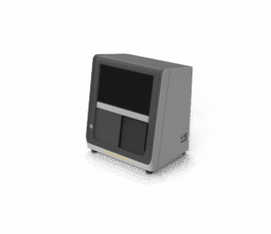
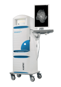
Kubtec PICASSO Plus
Specimen radiography · amorphous selenium · pathology gross room · Kubtec
View Product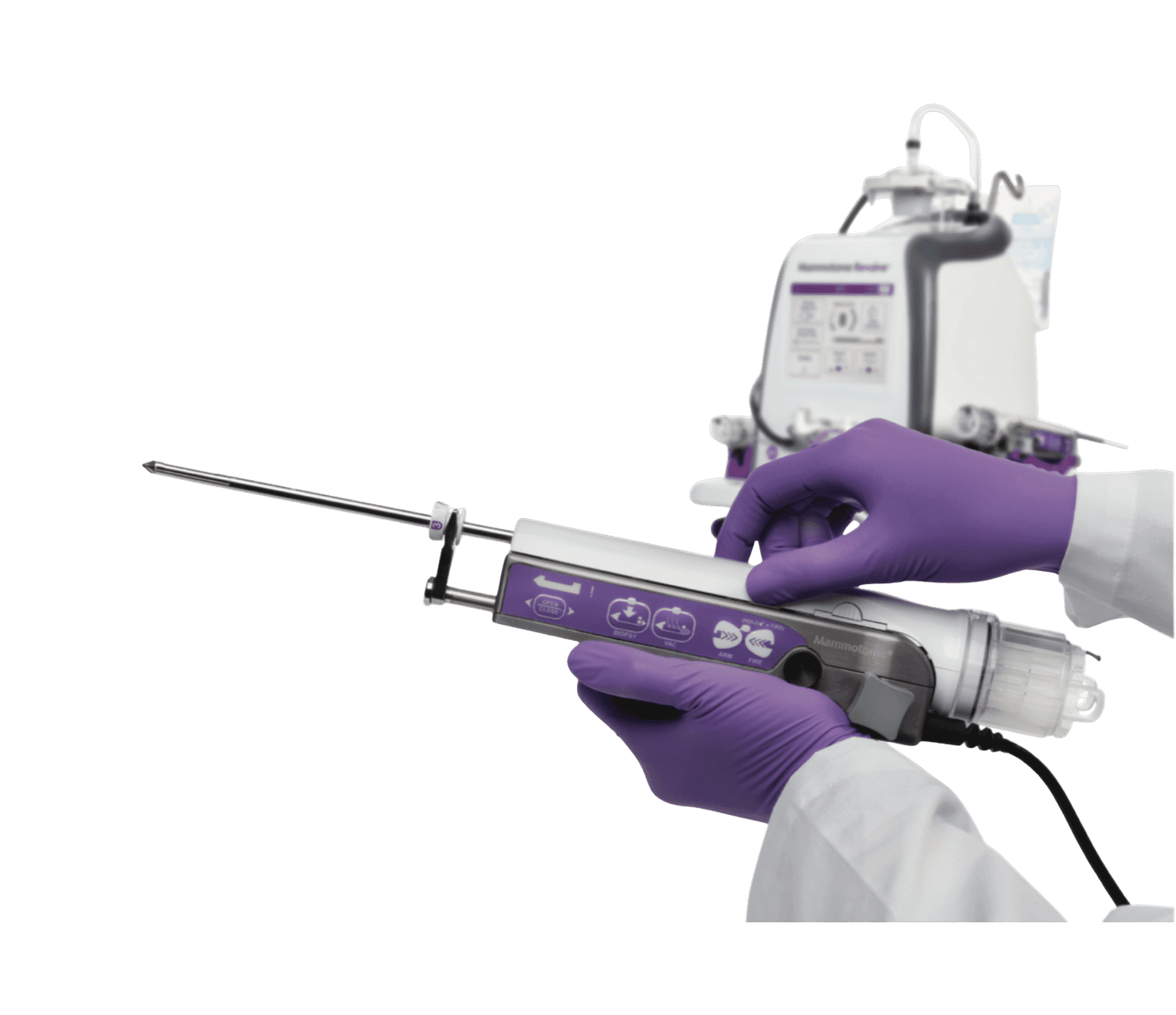
Mammotome Revolve
Vacuum assisted biopsy · breast tissue · image guided · pathology ready
View Product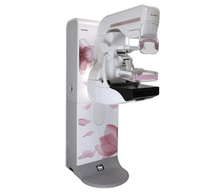
Fujifilm Aspire Cristalle
Digital mammography · tomosynthesis · breast imaging · low dose
View ProductRequest a Quote Today
Get in touch with Medical Outfitters today to request your personalized quote and outfit your facility with confidence.