
Koning Vera Breast CT overview
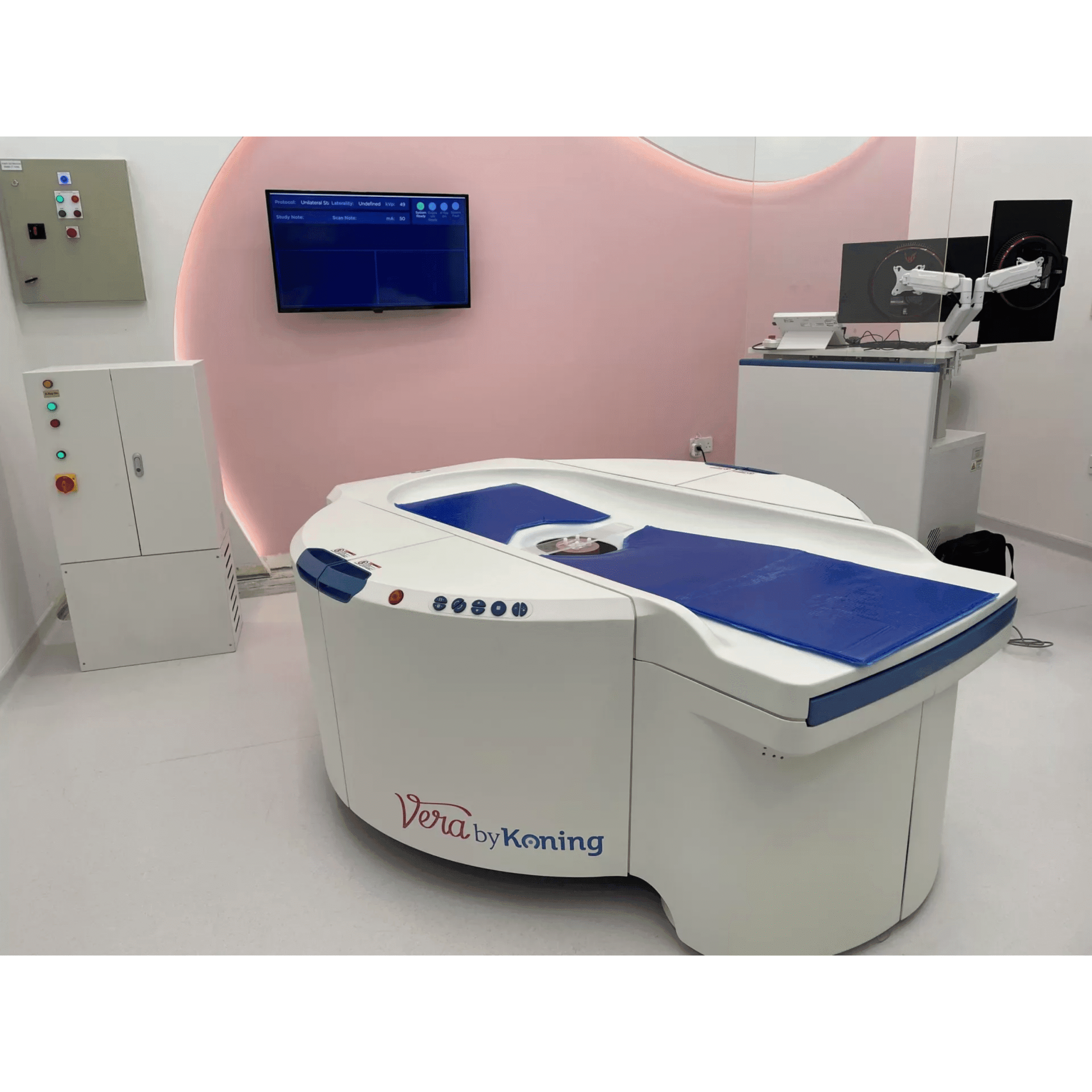
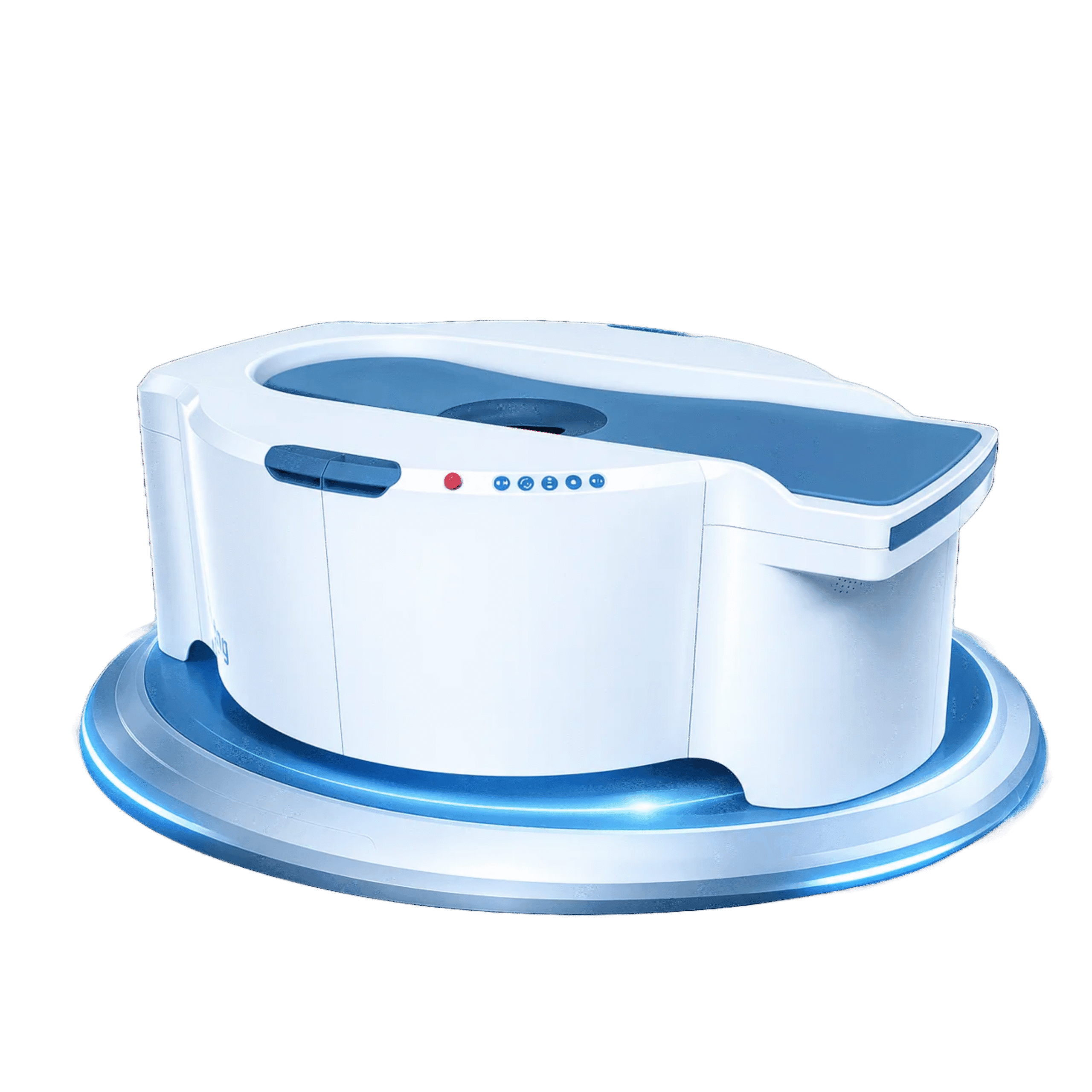
Authorized Koning Vera Breast CT supplier. The Koning Vera Breast CT is a dedicated cone-beam scanner built to image the entire breast from the chest wall to the nipple. It captures true 3D isotropic images in a single 10 second scan. As a result, structure and tissue overlap no longer obscures small lesions, and clinicians can detect tumors as small as 2 mm.
Traditional mammography is two-dimensional and requires patients to compress the breast with 10 to 20 kg of force. This scanner takes a different path. It positions the patient prone with no compression, so the exam is far more comfortable. In addition, its self-shielding design and dedicated operator console eliminate the need for a separate control room.
Need pricing? Get a quote with lead times within one business day.
Koning Vera Breast CT features
Koning has developed cone-beam CT technology for more than two decades and holds over 80 patents globally. Therefore the Vera Breast CT focuses on three goals: better detection, real patient comfort, and a workflow that fits existing rooms.
The system acquires a full 3D isotropic image in just 10 seconds. Because it captures 300 projections per scan, it builds volumetric anatomy without flattening the breast. Patients lie prone, and no compression force is applied. As a result, the procedure is comfortable while still detecting tumors as small as 2 mm.
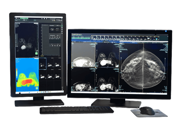
The self-shielding cabinet and dedicated operator console remove the need for a separate control room. Furthermore, the footprint fits standard stereotactic rooms, so costly construction is avoided. Every image is DICOM compliant. Therefore studies plug directly into most RIS and PACS systems for remote viewing and surgical planning.
Here is how the Vera Breast CT compares with conventional 2D screening mammography.
KBCT is the first commercially available CT scanner designed specifically to image the entire breast. Because it captures true 3D anatomy, it gives radiologists and breast surgeons a clear, overlap-free view of the tissue.
Full Specifications
All specifications below come from the Koning Breast CT System specification sheet. Values are listed as published by the manufacturer.
| Input option A | 480 V 3-phase @ 60 A plus ground |
| Input option B | 208 V 3-phase @ 120 A plus ground |
| Maximum voltage | 49 kVp |
| Maximum current | 200 mA |
| Power output | 9.8 kW |
| Temperature | 20° C to 24° C |
| Humidity | 30% to 60% rH, non-condensing |
| Minimum room size | 5.5 m × 6.0 m |
| Air Kerma | 25 mGy ±20% |
| Half value layer (FDA specification) | >0.49 mm Al at 49 kVp |
| Half value layer (typical) | 1.5 mm Al ±10% at 49 kVp |
| Scan time | 10 seconds |
| Projections | 300 per scan |
| Standard voxel | 0.273 mm isotropic |
| High resolution voxel | 0.190 mm and 0.155 mm isotropic |
| Patient table load | 200 kg maximum |
| Patient table height | 1.0 m to 1.55 m, ±10% |
| Patient position | Prone |
| Patient access | Both sides, interlocking safety covers |
Clinical Applications
The Vera Breast CT supports a range of breast imaging and care pathways. Here are six common scenarios.
Because the scanner removes tissue overlap, it helps reveal early-stage lesions. Therefore it supports detection of tumors as small as 2 mm.
Mammography often struggles with small dense breasts. In contrast, true 3D isotropic imaging captures clear detail in dense tissue.
Patients lie prone with no compression. As a result, a procedure many women dread becomes far more tolerable.
The table elevates to 1.55 m and accepts an optional biopsy kit. Therefore biopsies can be done on the table without a separate stereotactic system.
Breast surgeons use the full 3D anatomy to plan procedures. Because the volume is isotropic, it shows true spatial relationships.
Every study is DICOM compliant. As a result, radiologists view and share images remotely through most RIS and PACS systems.
Breast imaging workflow
The Vera Breast CT was designed to slide into existing clinical workflows. Each step below shows how the system supports the care team.
The patient lies prone on the dedicated exam table. No compression is applied at any point. A full 3D image is then acquired in about 10 seconds across 300 projections.
The console reconstructs true 3D isotropic volumes. Standard and high resolution voxels are available at 0.273 mm, 0.190 mm, and 0.155 mm. Therefore radiologists can scale detail to the clinical question.
The table elevates up to 1.55 m and wide safety covers open from both sides. With the optional biopsy kit, biopsies are performed directly on the table. As a result, a separate stereotactic biopsy table is not needed.
Images are DICOM compliant and connect directly to most RIS and PACS systems. Therefore reading, archiving, and remote review fit the existing enterprise workflow.
Breast surgeons receive full 3D anatomy for planning. Because the data is isotropic, it supports accurate spatial assessment ahead of the procedure.
Compliance and installation
The Vera Breast CT is built around interoperability, dose transparency, and a compact site footprint.
All images are DICOM compliant. Therefore they integrate with most RIS and PACS systems for remote viewing, archiving, and reporting.
The system delivers a radiation dose equivalent to mammography, with an Air Kerma of 25 mGy ±20%. The half value layer is greater than 0.49 mm Al at 49 kVp, which meets the published FDA specification.
The self-shielding design and dedicated console remove the need for a separate control room. In addition, the footprint fits standard stereotactic rooms, so costly construction is avoided.
Medical Outfitters Inc. is an ISO 13485:2016 certified medical imaging equipment supplier based in Miami, FL. Therefore every Koning Vera Breast CT order is handled under a documented, audited quality management system.
Request a quote
Contact our team for pricing, financing options, and lead times on the Koning Vera Breast CT. Financing ranges from lease buy-back to click-per-scan revenue sharing.
Installation and training
Technician training is provided with each install. In addition, customers gain access to a global medical advisory board, plus service and parts warranties offered for the life of the product.
Frequently Asked Questions
Common questions about the Koning Vera Breast CT scanner.
The system acquires a full 3D image in about 10 seconds. It captures 300 projections during that single scan.
No. The patient lies prone and no compression is applied. As a result, the procedure is far more comfortable than mammography.
Because true 3D isotropic imaging removes tissue overlap, the system supports detection of tumors as small as 2 mm.
Yes, with the optional biopsy kit. The table elevates up to 1.55 m, so a separate stereotactic biopsy table is not needed.
Yes. All images are DICOM compliant and plug directly into most RIS and PACS systems for remote viewing.
The minimum recommended room is 5.5 m by 6.0 m. It runs on 480 V 3-phase at 60 A, or 208 V 3-phase at 120 A, plus ground.
The dose is equivalent to mammography. The Air Kerma is 25 mGy ±20%, and the half value layer meets the published FDA specification.
You can buy it from Medical Outfitters Inc., an ISO 13485:2016 certified imaging equipment supplier in Miami, FL. Call (305) 885-4045 for pricing, financing, and installation.
Compatible Equipment
Explore related breast imaging and biopsy systems available from Medical Outfitters Inc.
Request a Quote Today
Get in touch with Medical Outfitters today to request your personalized quote and outfit your facility with confidence.


