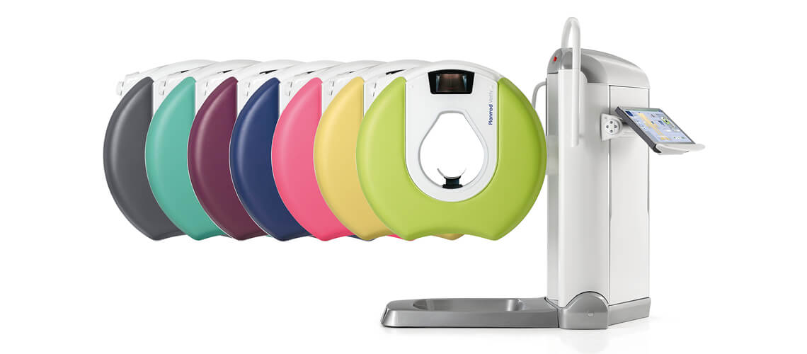
Planmed Verity: Weight-Bearing Extremity CBCT Scanner
Authorized Planmed extremity CBCT equipment supplier. The Planmed Verity is a cost-effective, space-saving extremity CT scanner designed for effective lower extremity exams, and with a few core modifications it can also be used for head and neck exams. It is the world’s first cone beam CT scanner designed for weight-bearing orthopedic imaging. Use the Verity to assess fractures and malunions, to evaluate the healing of fractures with no need for cast removal, and for preoperative planning.
The Verity is a compact, versatile CBCT that produces exceptional 3D images while exposing patients to a very low radiation dose. Weight-bearing imaging reveals issues like impingement that a typical exam can disguise, stitching algorithms combine images into a single view for extended exams, and true 3D vision with isotropic voxels and multiplanar reconstruction supports an accurate diagnosis. A flat panel sensor, a Multi field of view up to 16 by 20 cm, 200 to 400 micron voxels, Planmeca CALM movement correction, metal artifact removal, and a self-shielded portable design keep the workflow simple and the footprint small.
Need pricing? Get a quote with lead times and volume discounts within one business day.
Weight-Bearing Extremity 3D Imaging in a Compact CBCT
The Verity is a compact, versatile CBCT that produces exceptional 3D images at a very low dose. Weight-bearing capture, a TearDrop open gantry, and a self-shielded portable design set it apart for orthopedic extremity imaging. Full technical detail is in the expandable sections below.
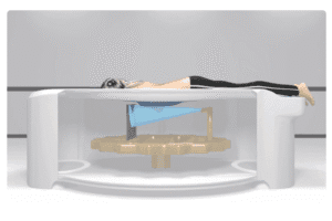
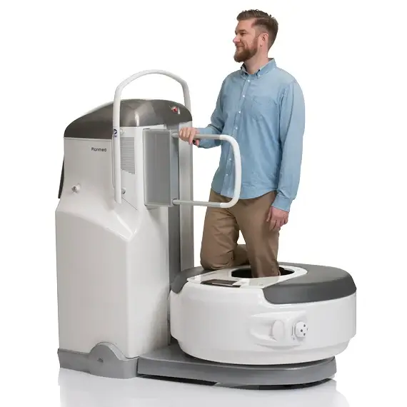
Weight-Bearing Imaging: the first CBCT of its kind to capture images in a natural weight-bearing position, tilting the gantry horizontal so the patient stands inside it to image the toes, ankles, feet, and knees under load
Reveal Hidden Findings: imaging under natural load highlights areas of contact and impingement that remain hidden or doubtful in a non-weight-bearing position, and seated and standing exams can be compared side by side
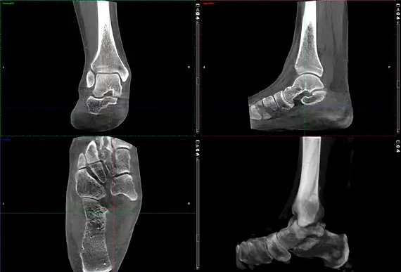
True 3D Vision: isotropic high-resolution voxels and multiplanar reconstruction show bone structures without overlapping anatomy for an accurate diagnosis
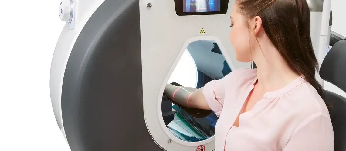
TearDrop Open Gantry: an extended TearDrop opening gives access from both sides of the gantry and reduces anxiety and claustrophobia, with red and green positioning lasers and a video camera
Dedicated Positioning: anatomically designed carbon fiber trays, a positioning camera, and a motorized gantry adjustable in height and tilt keep the target in the field of view
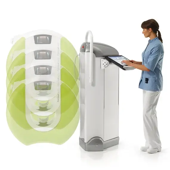
Stitching: a stitching algorithm combines images into a single volume for anatomies that need an extended imaging volume
Self-Shielded Mobility: a compact, portable design fits almost any radiology room with minimal shielding and can sit alongside existing equipment, needing only standard mains power and an Ethernet connection
Ultra Low Dose: the Planmeca Ultra Low Dose protocol delivers CBCT at a dose similar to a 2D X-ray extremity study and far below conventional MDCT, without reducing diagnostic quality
Planmeca CALM: movement artifact correction eliminates motion artifacts, which is invaluable for pediatric, weight-bearing, and complex patient exams
Metal Artifact Removal: an intelligent MAR algorithm maximizes the visibility of implants, fixators, and prostheses and the surrounding bone for post-operative imaging
OneScan and Reconstruction: OneScan positioning with automated correction avoids unnecessary retakes, and iterative 3D reconstruction refines the image in real time
Motorized Positioning: a motorized, one-hand control system positions the patient precisely so staff spend less time pushing and more time comforting
How the Verity compares against conventional MDCT for extremity imaging:
Up to 30 percent of scaphoid fractures can be missed on a 2D radiograph after trauma, and delayed diagnosis can lead to serious complications. The Verity brings high-resolution, low dose 3D imaging to the point of care for improved fracture diagnostics, and its weight-bearing capability reveals impingement and joint contact under natural load. Combined with metal artifact removal for post-operative imaging, the ability to assess fracture healing without removing a cast, and Disior 3D analytics for preoperative planning, the Verity changes orthopedic imaging for the better.
Full Technical Specifications
Complete reference data for capital procurement, RFP submissions, and biomedical review. Orthopedic surgeons, radiologists, and biomedical engineers rely on this specification set when they evaluate a dedicated extremity cone beam CT system. Expand each category for full detail.
| Modality | Flat panel cone beam CT for orthopedic extremity imaging |
| Power | 80 to 96 kV, 1 to 12 mA |
| Field of View | Multi field of view up to 16 by 20 cm |
| Voxel Sizes | 200 to 400 microns, isotropic |
| Reconstruction | Iterative 3D reconstruction in real time with multiplanar reconstruction and surface rendering |
| Upper Extremities | Elbow, arm, wrist, hand, and fingers |
| Lower Extremities | Knee, leg, ankle, foot, and toes, with weight-bearing capability |
| Head and Neck Option | Face, dental arch, TMJ, neck, sinuses, and teeth |
| Positioning Trays | Anatomically designed carbon fiber trays with a positioning camera |
| Gantry | Motorized open TearDrop gantry adjustable in height and tilt with one-hand control |
| Guidance | Red and green positioning lasers and a video camera on the gantry monitor |
| Ultra Low Dose | Planmeca Ultra Low Dose protocol for CBCT well below conventional MDCT |
| Comparable Dose | One scan at a dose similar to a 2D X-ray extremity study |
| Movement Correction | Planmeca CALM eliminates motion artifacts for pediatric and weight-bearing exams |
| Metal Artifact Removal | Intelligent MAR maximizes visibility of implants, fixators, and prostheses |
| OneScan | OneScan positioning with automated correction avoids unnecessary retakes |
| Artifact Removal | Advanced artifact removal up to the bone and metal interface |
| Dimensions | 76 by 184 by 160 cm, width by length by height |
| Weight | Approximately 350 kg |
| Power Input | 100 to 240 V single phase, 10 to 16 A |
| Shielding | Self-shielded with minimal shielding required, fits alongside existing equipment |
| Installation | Standard mains power and an Ethernet connection, no external cooling, portable |
| Verity Manager | Integrated image transfer and patient list software |
| DICOM | Modality Worklist, Storage, Query and Retrieve, Print, RDSR, and MPPS |
| Networks | Communicates with HIS, RIS, and PACS |
| Romexis Medical | Planmeca Romexis Medical for viewing, processing, and storing volumetric images |
| Disior Bonelogic | Optional 3D analytics for hand and wrist, foot and ankle, and orbital fractures |
| Accessories | Verity StackRack tray holder and an optional workstation |
Clinical Applications
Deployed across orthopedics and trauma where dedicated, low dose 3D extremity imaging under natural load improves fracture diagnostics, joint assessment, and surgical planning.
High-resolution 3D imaging at the point of care improves detection of fractures such as occult scaphoid fractures that 2D radiographs can miss.
Imaging the foot, ankle, and knee under natural load reveals impingement and joint contact that a non-weight-bearing exam can disguise.
Metal artifact removal maximizes visibility of implants, fixators, and prostheses and the bone around them for post-operative assessment.
The bone healing process can be evaluated without removing the cast, which has led to shorter immobilization times.
True 3D vision and Disior Bonelogic analytics provide objective measurements for patient-specific surgical planning.
Equipped for head and neck, the Verity can image the face, dental arch, TMJ, sinuses, and teeth for added versatility.
From Positioning to a 3D Analytics Report
An extremity imaging workflow built for low dose and simplicity. Tray-guided positioning, seated or weight-bearing capture, low dose CBCT, and 3D analytics move the case from setup to surgical planning. Expanded workflow details are in the expandable sections below.
The technician selects the anatomically designed carbon fiber positioning tray for the extremity and uses the positioning camera, red and green lasers, and the motorized one-hand control to center the target in the field of view. The extended TearDrop opening gives access from both sides of the gantry and reduces patient anxiety.
For routine exams the patient is seated, and for lower-joint assessment the gantry tilts to a horizontal position so the patient can stand inside it and be imaged under natural load. Seated and weight-bearing exams can be captured for side-by-side comparison.
The flat panel CBCT acquires the volume at 80 to 96 kV using the Planmeca Ultra Low Dose protocol, at a dose similar to a 2D X-ray extremity study. Planmeca CALM corrects patient movement, OneScan positioning avoids retakes, and metal artifact removal preserves visibility around implants.
Iterative 3D reconstruction refines the image in real time, and stitching extends the volume when needed. Volumetric data opens in Planmeca Romexis Medical and optional Disior Bonelogic analytics for objective measurements, and DICOM images archive automatically to PACS for review on any networked workstation.
Regulatory Compliance and Safety
FDA cleared as an extremity cone beam CT scanner, manufactured by Planmed Oy of Finland. Medical Outfitters is an ISO 13485:2016 certified supplier of imaging equipment, so every Planmed Verity ships through a documented and audited quality management system. Full compliance documentation is available in the expandable sections below.
FDA Cleared: the Planmed Verity extremity CBCT scanner is FDA cleared.
ISO 13485: Medical Outfitters Inc. is independently ISO 13485:2016 certified as a supplier, so every system ships through a fully audited quality management system.
Ultra Low Dose: the Planmeca Ultra Low Dose protocol keeps CBCT dose similar to a 2D X-ray extremity study and well below conventional MDCT.
Dose Reporting: the system supports radiation dose structured reports through DICOM for dose tracking and compliance.
Manufacturer: the Verity is manufactured by Planmed Oy of Helsinki, Finland.
Integration: full DICOM compatibility with Modality Worklist, Storage, Query and Retrieve, Print, and MPPS connects the unit to HIS, RIS, and PACS.
Configuration & Software Review
Tell us your orthopedic caseload and whether you need the head and neck option or Disior analytics. Our team confirms the configuration, positioning trays, and software, and arranges installation and training.
Capital Equipment & RFP Support
Pricing, technical documentation, and logistics for extremity CBCT deployments. We support orthopedic practices, imaging centers, and purchasing departments evaluating cone beam CT systems. Contact us for RFP packages.
Frequently Asked Questions
Quick answers to the most common purchase and clinical questions about the Planmed Verity extremity CBCT scanner. Tap any question to expand.
The Planmed Verity is the world’s first cone beam CT scanner designed for weight-bearing orthopedic imaging, dedicated to 3D extremity imaging. It captures high-resolution, low dose 3D images of the upper and lower extremities at the point of care, and it can also be equipped for head and neck imaging. It is used to assess fractures and malunions, evaluate fracture healing without cast removal, and support preoperative planning.
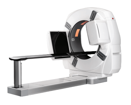
Both are Planmed orthopedic CBCT scanners, but they serve different scopes. The Verity is a dedicated extremity CBCT, compact, self-shielded, and portable, that images the upper and lower extremities and the lower joints under weight-bearing load with a Multi field of view up to 16 by 20 cm. The Planmed XFI is a larger full-body weight-bearing CBCT that also covers the spine and pelvis. Our team helps match the right system to your caseload and space.
The Verity images the upper extremities, including the elbow, arm, wrist, hand, and fingers, and the lower extremities, including the knee, leg, ankle, foot, and toes, with the lower joints imaged under weight-bearing load. Equipped for head and neck imaging, it can also scan the face, dental arch, TMJ, neck, sinuses, and teeth, which adds versatility and a fast return on investment.
Weight-bearing extremity CBCT images the lower joints while the patient stands under natural load. The gantry tilts to a horizontal position so the patient can stand inside it, which highlights areas of contact and impingement that would remain hidden in a non-weight-bearing position. Comparison exams between sitting and standing positions reveal changes in detail with high quality 3D images.
A flat panel CBCT scan uses a significantly lower radiation dose than conventional MDCT. One high-resolution Verity scan uses dose levels similar to those of a 2D X-ray extremity study, which corresponds to less than a week of naturally occurring background radiation. The Planmeca Ultra Low Dose protocol reduces the dose further without reducing diagnostic quality.
The Planmed Verity Manager software provides DICOM connectivity to the HIS, RIS, and PACS, with Modality Worklist, Storage, Query and Retrieve, Print, radiation dose structured reports, and MPPS. Volumetric images can be viewed and processed in Planmeca Romexis Medical, and Disior Bonelogic 3D analytics modules for hand and wrist, foot and ankle, and orbital fractures provide objective measurements for diagnosis and surgical planning.
Yes. The Planmed Verity extremity CBCT scanner is FDA cleared. Medical Outfitters Inc. is ISO 13485:2016 certified as the supplying distributor, so every system ships through an audited quality management system. It is manufactured by Planmed Oy of Helsinki, Finland.
Through Medical Outfitters Inc., ISO 13485:2016 certified, Miami, FL. We supply the system with positioning trays, the Verity Manager and Romexis Medical software, and optional Disior analytics, and provide installation and training. Call (305) 885-4045 or request a quote online.
Compatible Equipment
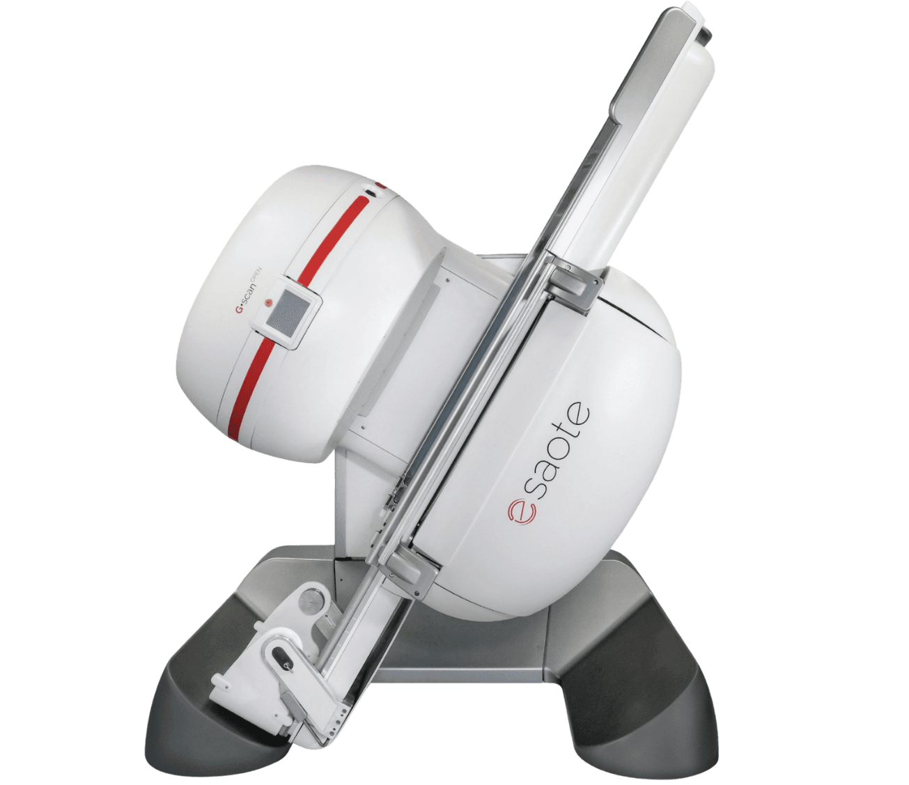
Other orthopedic imaging systems to consider alongside the Planmed Verity, from the Planmed XFI full-body weight-bearing CBCT to the Esaote G-scan Open weight-bearing MRI and the Orthoscan TAU 1515 mini C-arm.
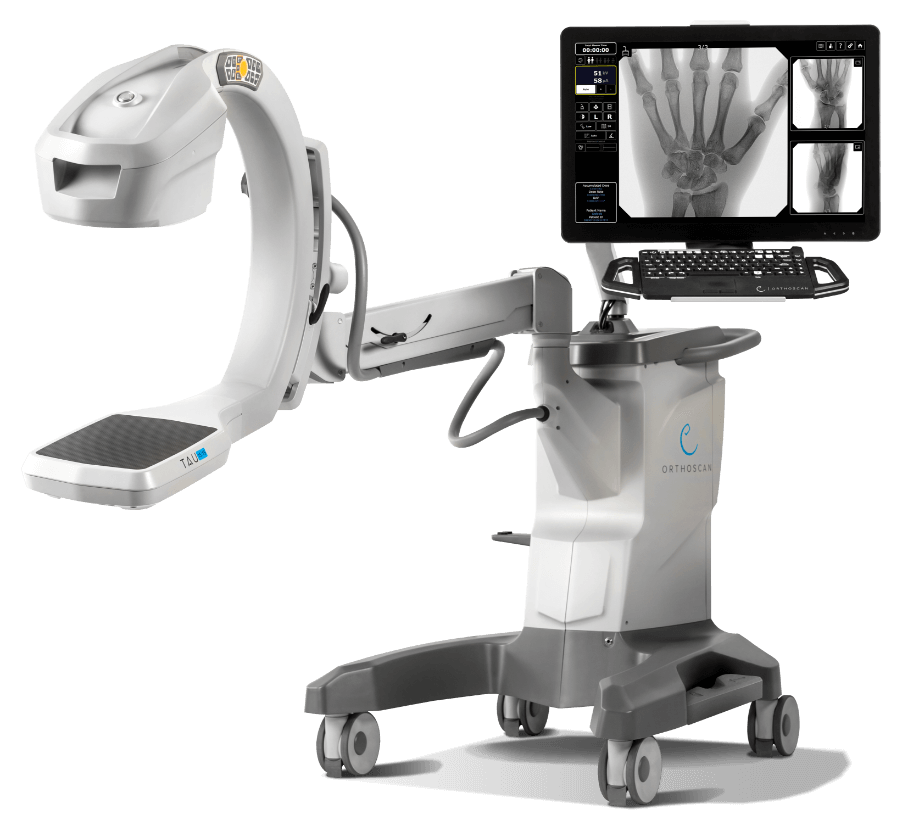
Orthoscan TAU 1515
Mini C-arm · extremity fluoroscopy · pediatric indication · Orthoscan
View ProductRequest a Quote Today
Get in touch with Medical Outfitters today to request your personalized quote and outfit your facility with confidence.