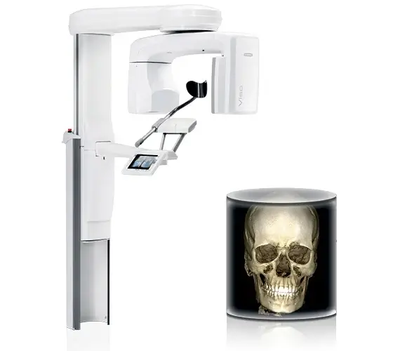
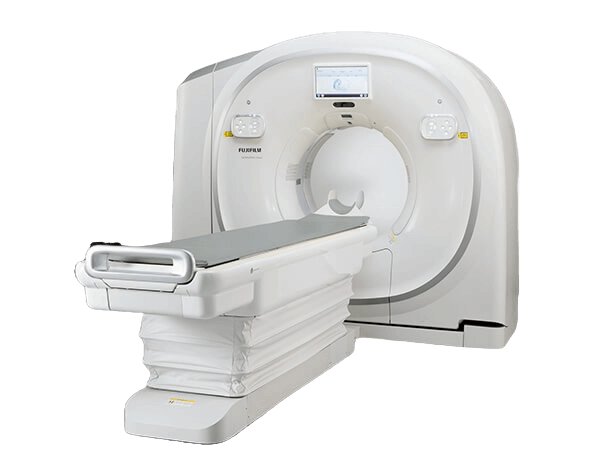
Planmeca Viso G7: 3D Dental and Maxillofacial CBCT
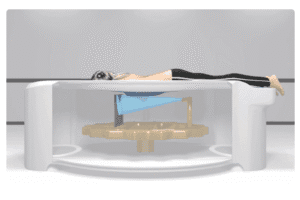
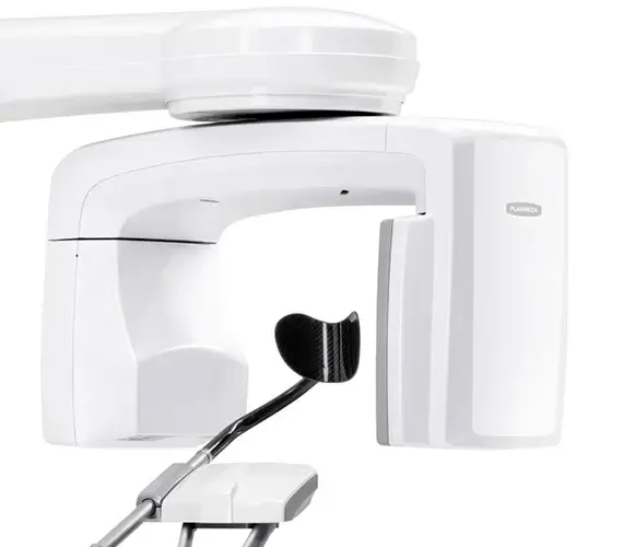
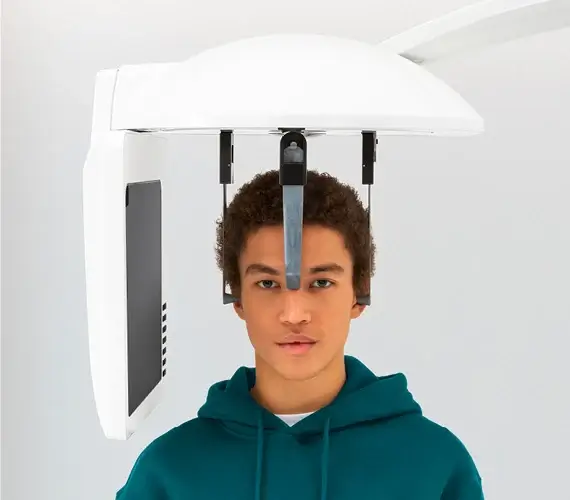
Authorized Planmeca dental CBCT equipment supplier. The Planmeca Viso G7 is a 3D cone beam CT imaging device designed to image the head and neck. It is packed with intelligent software that ensures a low radiation dose without sacrificing crisp images, and it is built for dentistry, orthodontics, endodontics, oral and maxillofacial surgery, and ENT applications. Patient positioning is performed directly from the control panel for easy alignment, and an open format with rear head and neck support keeps patients calm and comfortable.
The Viso G7 covers the entire maxillofacial area in a single scan, and the volume adjusts freely from about 3 by 3 cm to 30 by 30 cm with live virtual field of view positioning. A 60 to 120 kV flat panel CBCT captures high definition images down to a 75 micron endodontic voxel, while Planmeca Ultra Low Dose imaging and Planmeca CALM movement artifact correction keep the dose low and the images sharp. Planmeca ProFace 3D face photography, preprogrammed dental, orthodontic, and ENT programs, and Planmeca Romexis integration round out the system.
If you are looking for top-tier CBCT imaging with exceptional detail and a flexible field of view, the Viso G7 is a standout. It is well suited to everything from endodontics to airway analysis.
Need pricing? Get a quote with lead times and volume discounts within one business day.
Dynamic Volumes, Ultra Low Dose, and ProFace 3D Imaging
The Viso G7 is designed for practices that demand premium image quality without compromising patient comfort or workflow efficiency. Dynamic volumes, live positioning, low dose imaging, and ProFace 3D photos set it apart. Full technical detail is in the expandable sections below.
Dynamic Volume Selection: a wide volume selection covers areas as small as a single tooth and as large as the entire skull, freely adjustable from about 3 by 3 cm up to 30 by 30 cm
Single-Scan Maxillofacial Coverage: the entire maxillofacial area can be captured in a single scan up to about 30 by 19 cm
Live Patient Positioning: patient positioning is performed from the control panel using integrated cameras, with the patient always facing the operator to reduce awkwardness and motion
Live Virtual FOV Positioning: the operator adjusts the field of view on screen with the tip of a finger before the scan
Advanced Touchscreen Interface: a graphical user interface with preprogrammed settings is quick and easy for anyone to use, including staff with little imaging experience
Open, Comfortable Design: an open format with rear head and neck support keeps patients calm, which reduces retakes and improves comfort
Planmeca Ultra Low Dose: a proprietary protocol uses algorithms to calculate the lowest dose for a given anatomical region and exam type, works with all voxel sizes and modes, and displays the dose on the touchscreen
High Definition Voxel Imaging: small voxel sizes from 600 microns down to a 75 micron endodontic mode produce sharp, accurate 3D images for implant planning and endodontics
Planmeca CALM Movement Correction: movement artifact correction compensates for patient movement to minimize artifacts, even when patients do not stay still
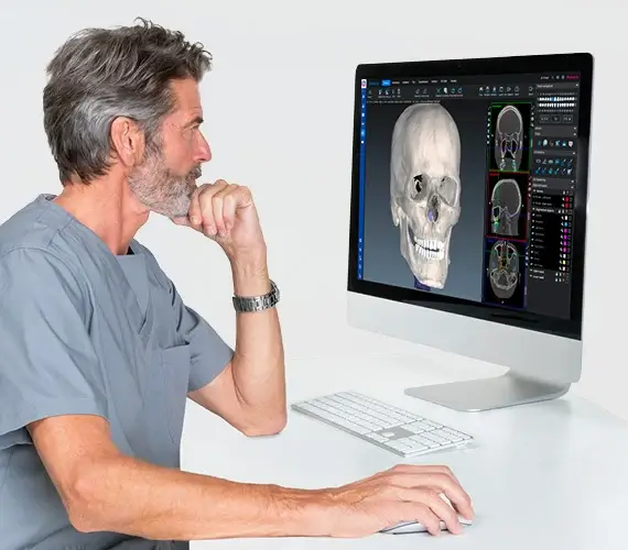
Planmeca ProFace 3D Face Photo: four cameras and an LED light capture realistic 3D facial photographs that combine with scans to enrich treatment plans
Planmeca Romexis Integration: the Viso G7 integrates with Planmeca Romexis software and the mRomexis tablet application for streamlined workflows
DICOM Support: DICOM standard compliance ensures the unit works with most existing and legacy systems
How the Viso G7 compares against a conventional fixed field of view dental CBCT:
The Viso G7 adapts to the clinical question rather than forcing the case into a fixed field of view. The operator selects exactly the volume needed, from a 3 by 3 cm endodontic scan at a 75 micron voxel to a full 30 by 30 cm view of the skull, and positions the patient live from the control panel. Planmeca Ultra Low Dose keeps exposure to the minimum required, CALM corrects for movement, and ProFace adds a photorealistic 3D face layer for richer treatment planning and patient communication.
Full Technical Specifications
Complete reference data for capital procurement, RFP submissions, and biomedical review. Dentists, oral and maxillofacial surgeons, orthodontists, and biomedical engineers rely on this specification set when they evaluate a dental cone beam CT system. Expand each category for full detail.
| Modality | 3D dental and maxillofacial cone beam CT |
| Anode Voltage | 60 to 120 kV |
| Anode Current | 1 to 16 mA |
| Focal Spot | 0.5 mm, fixed anode |
| Image Detector | Flat panel |
| Image Acquisition | 200 or 360 degree rotation |
| Scan Time | 1 to 36 seconds, reconstruction 2 to 55 seconds |
| Single Scan Volume | Up to about 30 by 19 cm covering the entire maxillofacial area |
| Maximum Volume | About 30 by 30 cm |
| Minimum Volume | About 3 by 3 cm |
| Dynamic Selection | Freely adjustable from a single tooth to the entire skull |
| Voxel Sizes | 600 and 400 microns low dose, 200 micron Normal, 150 micron High Definition and Braces, 100 micron High Resolution, 75 micron Endodontic |
| Ultra Low Dose | Planmeca Ultra Low Dose protocol for all voxel sizes and modes, with on-screen dose display |
| Movement Correction | Planmeca CALM movement artifact correction |
| High Definition | Small voxel imaging for precise implant and endodontic planning |
| Dose Tracking | X-ray log book records exposure values including kV and mAs |
| 3D Dental Programs | Preprogrammed dental imaging programs |
| 3D ENT Programs | Ear, nose, and throat imaging programs |
| Endodontic Mode | Best resolution at the smallest 75 micron voxel size |
| 2D Imaging | 2D panoramic imaging and one-shot cephalometric imaging |
| Motion and Models | 4D jaw motion and 3D models scan |
| Clinical Uses | Implant planning, endodontics, orthodontics, oral surgery, and airway analysis |
| Planmeca ProFace | Optional 3D face photo system with four cameras and an LED light |
| Live Positioning | Patient positioning from the control panel with integrated cameras |
| Romexis | Planmeca Romexis software integration and the mRomexis tablet application |
| RFID | Integrated RFID capabilities |
| Connectivity | DICOM standard compliant for use with most existing and legacy systems |
Clinical Applications
Deployed across dentistry, oral and maxillofacial surgery, orthodontics, endodontics, and ENT where adjustable 3D cone beam imaging of the head and neck guides diagnosis and treatment planning.
High definition small voxel imaging supports precise dental implant planning and placement.
Endodontic mode delivers the best resolution at a 75 micron voxel for detailed root canal and periapical assessment.
Dynamic volume selection and a one-shot cephalometric option support orthodontic diagnosis and treatment planning.
3D ENT programs and large field of view scans support imaging of the sinuses, ear bones, and airway analysis.
Single-scan maxillofacial coverage and ProFace 3D photos support surgical planning and patient communication.
4D jaw motion captures dynamic movement of the lower jaw for temporomandibular joint assessment.
From Program Selection to a Romexis 3D Model
A dental imaging workflow built for comfort and precision. Program and volume selection, live positioning, low dose acquisition, and Romexis integration move the case from setup to treatment planning. Expanded workflow details are in the expandable sections below.
The operator selects a preprogrammed dental, orthodontic, or ENT program from the advanced touchscreen interface, then adjusts the field of view live on screen with a fingertip. The volume is set to exactly what the case needs, from a 3 by 3 cm endodontic scan to a full 30 by 30 cm view of the skull.
Patient positioning is performed from the unit control panel using integrated cameras, with the patient seated in an open format facing the operator and supported at the rear of the head and neck. This keeps the patient calm and still, which reduces repeat imaging and improves comfort.
The 60 to 120 kV flat panel CBCT acquires the volume with a 200 or 360 degree rotation in as little as one second. Planmeca Ultra Low Dose calculates the lowest dose for the region and exam type and displays it on screen, while Planmeca CALM corrects for patient movement to keep the image sharp.
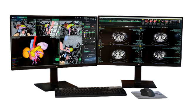
The volume reconstructs quickly and opens in Planmeca Romexis software, where it can be combined with a ProFace 3D face photo for a layered, photorealistic model. DICOM compliance and the mRomexis tablet application let the team share and view images across existing systems and devices.
Regulatory Compliance and Safety
FDA cleared as a dental cone beam CT imaging system, manufactured by Planmeca Oy of Finland. Medical Outfitters is an ISO 13485:2016 certified supplier of imaging equipment, so every Planmeca Viso G7 ships through a documented and audited quality management system. Full compliance documentation is available in the expandable sections below.
FDA Cleared: the Planmeca Viso G7 dental cone beam CT imaging system is FDA cleared.
ISO 13485: Medical Outfitters Inc. is independently ISO 13485:2016 certified as a supplier, so every system ships through a fully audited quality management system.
Ultra Low Dose: the Planmeca Ultra Low Dose protocol calculates and applies the lowest dose required for the region and exam type, with the dose displayed on the touchscreen.
Dose Logging: an X-ray log book automatically records image exposure values including kV and mAs to meet regulatory requirements.
Manufacturer: the Viso G7 is manufactured by Planmeca Oy of Helsinki, Finland.
Integration: the system is DICOM standard compliant and integrates with Planmeca Romexis software and the mRomexis tablet application for use with most existing and legacy systems.
Configuration & Software Review
Tell us your specialty mix across dentistry, orthodontics, and ENT and whether you need the ProFace option. Our team confirms the configuration and Romexis software, and arranges installation and training.
Capital Equipment & RFP Support
Pricing, technical documentation, and logistics for dental CBCT deployments. We support dental and oral surgery practices, imaging centers, and purchasing departments evaluating cone beam CT systems. Contact us for RFP packages.
Frequently Asked Questions
Quick answers to the most common purchase and clinical questions about the Planmeca Viso G7 dental cone beam CT imaging system. Tap any question to expand.
The Planmeca Viso G7 is a 3D dental and maxillofacial cone beam CT imaging system for the head and neck. It captures the entire maxillofacial area in a single scan, offers a freely adjustable field of view, and uses intelligent software to keep the radiation dose low without sacrificing crisp images. It is built for dentistry, orthodontics, endodontics, oral and maxillofacial surgery, and ENT applications.
Both are cone beam CT systems, but they serve different specialties and are different products. The Planmeca Viso G7 is a dental and maxillofacial CBCT for the head and neck with ProFace 3D face photography, dental and orthodontic programs, and an endodontic mode. The Planmed XFI is a full-body weight-bearing CBCT for orthopedic imaging. Our team helps match the right system to your specialty.
The Viso G7 covers the entire maxillofacial area in a single scan up to about 30 by 19 cm, and the volume size adjusts freely from about 3 by 3 cm up to 30 by 30 cm, so the team can image a single tooth or the whole skull. Voxel sizes range from 600 and 400 microns in low-dose modes, to 200 microns in Normal mode, 150 microns in High Definition and Braces modes, 100 microns in High Resolution, and 75 microns in Endodontic mode.
Planmeca Ultra Low Dose is a proprietary imaging protocol that uses algorithms to calculate the lowest dose required for a specific anatomical region and exam type, and it works with all voxel sizes and imaging modes, with the dose displayed on the touchscreen. Planmeca CALM is movement artifact correction that compensates for patient movement to minimize artifacts, which is especially valuable at small voxel sizes such as in endodontic imaging.
Planmeca ProFace is an exclusive 3D face photo system. The Viso G7 sensor uses four cameras and an LED light to capture realistic 3D facial photographs that can be combined with the CBCT and other scans to create a layered, photorealistic model. This enriches treatment planning and patient communication, and ProFace photos can also support simulation of treatment or surgery effects.
The Viso G7 includes preprogrammed 3D dental programs, 3D ENT programs, an endodontic mode, 2D panoramic imaging, one-shot cephalometric imaging, 3D models scan, and 4D jaw motion. This supports a wide range of uses including implant planning, endodontics, orthodontics, oral and maxillofacial surgery, airway analysis, and temporomandibular joint assessment.
Yes. The Viso G7 is integrated with Planmeca Romexis software for streamlined dental imaging workflows, and the Planmeca mRomexis tablet application lets you view images on a local network or carry them on a tablet. The system is DICOM standard compliant, so it can be used with most existing and legacy systems.
Through Medical Outfitters Inc., ISO 13485:2016 certified, Miami, FL. We supply the system with its software and accessories, including the ProFace option, and provide installation and training. Call (305) 885-4045 or request a quote online.
Compatible Equipment
Other 3D and cross-sectional imaging systems to consider alongside the Planmeca Viso G7, from the Planmed XFI weight-bearing cone beam CT to whole-body CT and advanced 3D visualization software.
Request a Quote Today
Get in touch with Medical Outfitters today to request your personalized quote and outfit your facility with confidence.