Planmed XFI: Full-Body Weight-Bearing Cone Beam CT
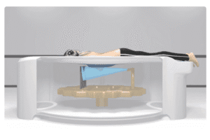
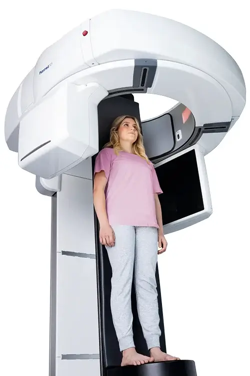
Authorized Planmed CBCT equipment supplier. The Planmed XFI is a unique cone beam computed tomography system that provides comprehensive imaging in both standing weight-bearing and supine positions. A wide opening and a broad field of view let the team capture the patient under natural physiological load, and operators can perform supine or weight-bearing exams quickly and switch between them as needed. It is a cutting edge orthopedic imaging system designed for detailed, full-body weight-bearing assessments.
The Planmed XFI pairs a wide 85 cm opening with a large 43 by 43 cm flat panel detector and a high power X-ray source that rotates 360 degrees around the target, rapidly capturing detailed 3D views. It delivers ultra high resolution isotropic images up to 75 microns for exceptional clarity, while intelligent CBCT and image optimization technologies follow the ALADA principle to keep effective patient dose low. A motorized patient table, laser guided positioning, and a compact footprint make it easy to integrate into most radiology departments.
Need pricing? Get a quote with lead times and volume discounts within one business day.
Full-Body Weight-Bearing 3D Imaging with a Low Dose
The Planmed XFI is built around weight-bearing assessment, high resolution 3D volume, and low patient dose. An open bore, a large flat panel detector, and intelligent dose optimization set it apart from conventional CT. Full technical detail is in the expandable sections below.
Full-Body Weight-Bearing Imaging: capture comprehensive 3D images in standing weight-bearing and supine positions, and compare both exams for a more complete diagnostic picture
High Resolution 3D Volume: detailed isotropic 3D images up to 75 microns reveal small fractures, lesions, joint space narrowing, and other findings with clarity and precision
Imaging Under Natural Load: 3D weight-bearing imaging overcomes projection differences and overlapping structures to show anatomy in its natural, functional position
Wide Open 85 cm Bore: an open design with a 360 degree rotating source keeps patients comfortable, less likely to move, and easy to position
Broad Field of View: an extensive field of view images large anatomy in a single exam without exposing the patient to excess radiation
Low Dose CBCT Engine: intelligent CBCT and image optimization technologies follow the ALADA principle to deliver low effective patient doses with faster scanning
Automatic Exposure Control: an 80 to 140 kV generator with Automatic Exposure Control adapts the exposure to the anatomy for consistent quality
Motion and Metal Handling: image optimization helps correct for patient motion and reduce streaks and shadows caused by metal implants for clearer surrounding tissue
Optimal Positioning: a motorized patient table and laser guided positioning let operators set the correct angle and verify alignment before the exam begins
Compact Footprint: a relatively compact system integrates into most existing radiology departments without extensive remodeling or construction
PACS and DICOM Integration: full DICOM 3.0 compatibility with RIS and PACS integration makes it easy to upload, share, and save each exam
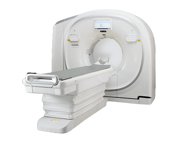
How the Planmed XFI compares against a conventional supine CT scanner:
Weight-bearing imaging plays a vital role in assessing musculoskeletal conditions, especially in the lower back and pelvis, by revealing pathologies that may not be visible when the body is at rest. The Planmed XFI clarifies spinal alignment, pelvic orientation, joint behavior under load, and the position of previously placed implants, and it enhances the understanding of biomechanical function. Integrating weight-bearing imaging into pre-surgical evaluation improves surgical decision making, reduces the risk of future complications, and supports better treatment outcomes.
Full Technical Specifications
Complete reference data for capital procurement, RFP submissions, and biomedical review. Radiology directors, orthopedic and spine surgeons, and biomedical engineers rely on this specification set when they evaluate a weight-bearing cone beam CT system. Expand each category for full detail.
| Detector | Large flat panel detector, 43 by 43 cm |
| Resolution | Up to 75 microns |
| Resolution Type | Isotropic resolution |
| Source to Image Distance | 108 cm |
| Rotation | 360 degree rotation of source and detector |
| kV Range | 80 to 140 kV |
| mA Range | 5 to 100 mA |
| Exposure Control | Automatic Exposure Control, AEC |
| Dose Principle | ALADA, As Low As Diagnostically Acceptable |
| Low Dose Technology | Intelligent CBCT and image optimization for low effective patient dose |
| Image Optimization | Motion handling and metal artifact reduction for clearer surrounding tissue |
| Image Quality | Low effective dose without compromising image quality |
| Bore | 85 cm opening |
| Field of View | Up to 23 by 44 cm |
| Dimensions | 248 by 176 by 162 cm, or 97.6 by 69.3 by 63.8 inches, length by height by width |
| Weight | Approximately 550 kg |
| Patient Table | Motorized patient table |
| Positioning | Laser guided positioning |
| Control Station | Remote control and acquisition workstation (AWS) |
| Connectivity | DICOM 3.0 compatibility with RIS and PACS integration |
| Line Voltage | 180 to 240 V, 50 Hz |
| Line Current | 16 A |
Clinical Applications
Deployed across orthopedics and spine care where imaging the body under natural load with low dose 3D cone beam CT changes the diagnosis and the surgical plan.
3D weight-bearing imaging clarifies spinal alignment and pelvic orientation, revealing pathologies in the lower back and pelvis that are not visible at rest.
Imaging the foot, ankle, knee, and hip under natural load shows joint behavior and alignment that supine CT cannot capture.
Functional, loaded 3D views support surgical decision making and reduce the risk of future complications in pre-surgical evaluation.
Assess previously placed implants under load to anticipate complications and confirm position after surgery.
Loaded imaging demonstrates joint space narrowing and osteoarthritic change that may be missed with conventional methods.
When a supine study is preferred, the same system captures high resolution isotropic 3D images of the extremities at a low dose.
From Loaded Positioning to a Low Dose 3D Volume
A weight-bearing CBCT workflow built for orthopedic assessment. Laser guided positioning, a fast 360 degree scan, low dose 3D reconstruction, and PACS integration move the case from scan to surgical decision. Expanded workflow details are in the expandable sections below.
The patient is positioned standing in a weight-bearing position, supine, or seated, depending on the clinical question. A motorized patient table and laser guided positioning let the operator set the correct angle and verify alignment before the exam, while the wide open 85 cm bore keeps the patient comfortable and still.
The large 43 by 43 cm flat panel detector and high power X-ray source rotate 360 degrees around the target, rapidly capturing the volume in a single exam. An 80 to 140 kV generator with Automatic Exposure Control adapts the exposure to the anatomy for consistent image quality.
Intelligent CBCT and image optimization reconstruct ultra high resolution isotropic 3D images up to 75 microns while following the ALADA principle to keep effective patient dose low. Image optimization helps correct for motion and reduce metal artifacts for clearer surrounding tissue.
The team can compare weight-bearing and supine exams for a more complete diagnostic picture, then route the results to PACS. Full DICOM 3.0 compatibility with RIS and PACS integration makes it easy to upload, share, and archive each study.
Regulatory Compliance and Safety
FDA 510(k) cleared under K250318 with CE mark pending, manufactured by Planmed Oy of Finland. The Planmed XFI is not cleared for imaging the head in the United States. Medical Outfitters is an ISO 13485:2016 certified supplier of imaging equipment, so every Planmed XFI ships through a documented and audited quality management system. Full compliance documentation is available in the expandable sections below.
FDA Cleared: the Planmed XFI is FDA 510(k) cleared under K250318, with CE mark approval pending.
Indication Limit: the Planmed XFI is not cleared for imaging the head in the United States.
ISO 13485: Medical Outfitters Inc. is independently ISO 13485:2016 certified as a supplier, so every system ships through a fully audited quality management system.
Manufacturer: the Planmed XFI is manufactured by Planmed Oy of Helsinki, Finland.
Configurations: images may contain optional items not included in standard delivery, and available configurations and features may have country or area specific variations.
Dose Management: the system follows the ALADA principle with intelligent CBCT, image optimization, and Automatic Exposure Control to keep effective patient dose low.
Connectivity: DICOM 3.0 compatibility with RIS and PACS integration.
Electrical: line voltage 180 to 240 V at 50 Hz with a 16 A line current.
Site Planning & Installation Review
Tell us your room dimensions and orthopedic caseload. Our team confirms the footprint and electrical fit, the weight-bearing and supine configuration, and coordinates installation and training.
Capital Equipment & RFP Support
Pricing, technical documentation, and logistics for cone beam CT deployments. We support radiology directors, orthopedic and spine surgeons, and purchasing departments evaluating weight-bearing CT systems. Contact us for RFP packages.
Frequently Asked Questions
Quick answers to the most common purchase and clinical questions about the Planmed XFI weight-bearing cone beam CT system. Tap any question to expand.
The Planmed XFI is a full-body weight-bearing cone beam computed tomography system, also called CBCT, designed for orthopedic imaging. It captures detailed 3D images in both standing weight-bearing and supine positions, using a wide 85 cm opening, a large 43 by 43 cm flat panel detector, and a high power X-ray source that rotates 360 degrees around the patient, with resolution up to 75 microns.
Weight-bearing CBCT images the body under its natural physiological load rather than only lying down. This reveals musculoskeletal pathologies that may not be visible at rest, particularly in the lower back and pelvis, and it clarifies spinal alignment, pelvic orientation, joint behavior under load, and the position of previously placed implants. The result supports more informed pre-surgical planning and better treatment outcomes.
A conventional CT scanner uses a fan beam helical design and images the patient only in a supine position inside a detector ring. The Planmed XFI is a cone beam CT with a large flat panel detector that scans in both standing weight-bearing and supine positions, captures isotropic 3D images up to 75 microns through an open 85 cm bore, and is purpose built for orthopedic assessment under load.
The Planmed XFI uses a large 43 by 43 cm flat panel detector with a source to image distance of 108 cm and delivers ultra high resolution isotropic 3D images up to 75 microns. It has a wide 85 cm bore and a field of view up to 23 by 44 cm, which captures detailed anatomy while the source and detector rotate 360 degrees around the target.
The Planmed XFI follows the ALADA principle, As Low As Diagnostically Acceptable, using intelligent CBCT and image optimization technologies to achieve low effective patient doses without compromising image quality. An 80 to 140 kV generator with Automatic Exposure Control adapts the exposure to the anatomy, which is especially important for children and for repeat assessments.
Patients can be scanned standing in a weight-bearing position, supine, or seated, and the team can switch between supine and weight-bearing exams to compare results. A motorized patient table and laser guided positioning make it easy for operators to set the correct angle and verify alignment before the exam begins.
Yes. The Planmed XFI is FDA 510(k) cleared under K250318, with CE mark approval pending. It is not cleared for imaging the head in the United States. Medical Outfitters Inc. is ISO 13485:2016 certified as the supplying distributor, so every system ships through an audited quality management system.
Through Medical Outfitters Inc., ISO 13485:2016 certified, Miami, FL. We supply systems with nationwide installation support, site planning, and training. Call (305) 885-4045 or request a quote online.
Compatible Equipment
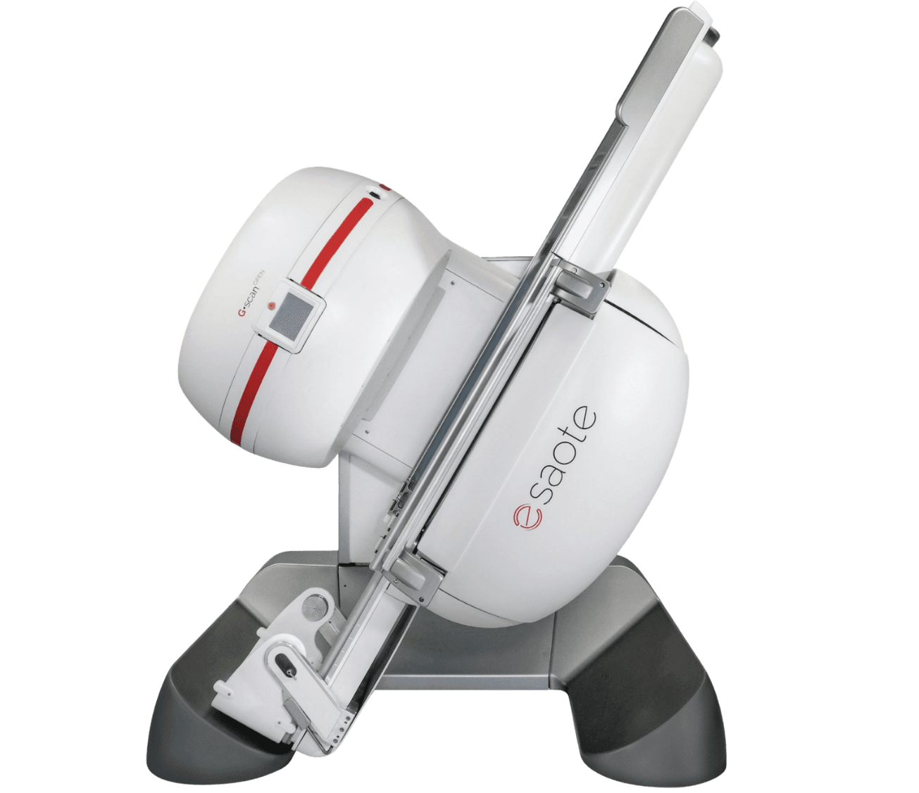
Imaging systems used alongside the Planmed XFI across the orthopedic pathway, from weight-bearing MRI and dedicated extremity imaging to mini C-arm fluoroscopy and whole-body CT.
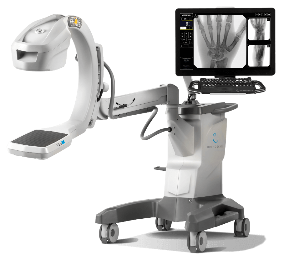
Orthoscan TAU 1515
Mini C-arm · extremity fluoroscopy · pediatric indication · Orthoscan
View ProductRequest a Quote Today
Get in touch with Medical Outfitters today to request your personalized quote and outfit your facility with confidence.