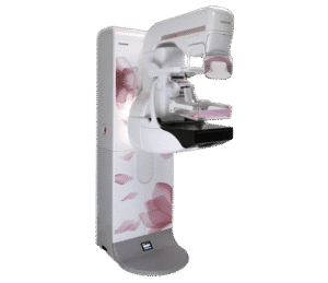
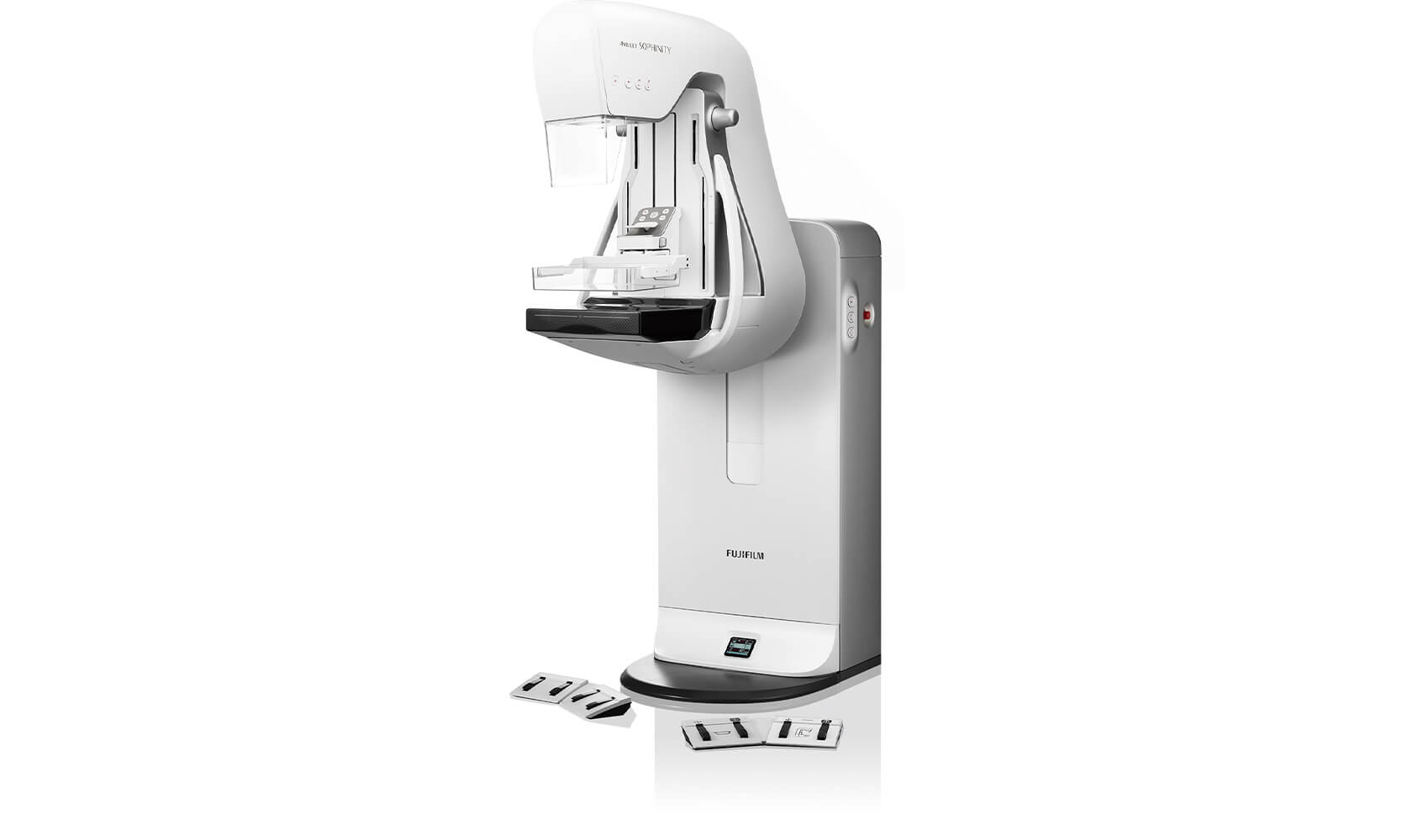
Fujifilm Amulet Sophinity: Digital Mammography System
Authorized Fujifilm breast imaging equipment supplier. The Amulet Sophinity is a digital mammography system that pairs high-resolution imaging with patient comfort at the center of its design. Developed by Fujifilm in close collaboration with patients, doctors, and technologists, it produces reliable diagnostic images at a low X-ray dose while making the exam experience smoother for everyone in the room. This is a 3D mammogram machine engineered for comfort, throughput, and diagnostic confidence, and a sound investment for facilities weighing mammogram cost against long-term screening volume.
Core breast imaging technology: the smallest direct-conversion 50 micron hexagonal pixel detector with a Tungsten target and ISC for high-definition microcalcification imaging at low dose. Optional digital breast tomosynthesis in Standard and High Resolution modes, S-View synthesized 2D, and CEDM contrast-enhanced mammography extend its diagnostic reach. Intelligent AEC, Comfort Comp automatic decompression, and AI-driven positioning support round out a digital mammography machine built to reduce pain and rescans alike.
Need pricing? Get a quote with lead times and volume discounts within one business day.
50 Micron Detail, Tomosynthesis & Comfort Design
Three pillars set the Amulet Sophinity apart from conventional digital mammography machines: high-definition detection, 3D tomosynthesis with synthesized 2D, and a comfort-first physical design. Full technical detail is in the expandable sections below.
50 Micron Hexagonal Pixel: The smallest direct-conversion FPD pixel size of 50 microns, arranged in a hexagonal pattern, enables high-definition imaging of microcalcifications that conventional square-pixel detectors may miss
Tungsten Target with ISC: Image-based Spectrum Conversion adjusts contrast while keeping the X-ray dose low using a Tungsten target
Dynamic Visualization: Density and contrast adjustment, frequency enhancement, and dynamic range compression automatically tune each image, recognizing mammary gland and fat areas and increasing contrast independently for consistent density
Multi-Frequency Processing (MFP): Optimizes fine structure and overall contrast across the full image
Microcalcification Clarity: The high-definition detector captures defects in crisp detail to give the diagnostic team more information per exam
Digital Breast Tomosynthesis: Continuous tube motion captures multiple projection images that are reconstructed into focused tomographic slices, reducing artifacts and revealing lesions hidden by overlapping mammary gland structures
Standard (ST) Mode: Plus or minus 7.5 degree sweep angle, 19 shots, 100 or 150 micron pixels, approximately 5 seconds, with deep depth of field for exams, screening, and follow-up
High Resolution (HR) Mode: Plus or minus 20 degree sweep angle, 35 shots, 50 or 100 micron pixels, approximately 12 seconds, with improved depth resolution and shallow depth of field for close examination
S-View Synthesized 2D: A synthetic 2D image including breast thickness information is generated from the tomosynthesis acquisition at 50 microns, so one session produces both a 3D image and a synthesized 2D image
CEDM Contrast Mammography: Low-energy and high-energy imaging with a Cu filter in a single compression automatically generates an energy subtraction image when an iodinated contrast medium is used
How the Amulet Sophinity compares against a conventional digital mammography machine:
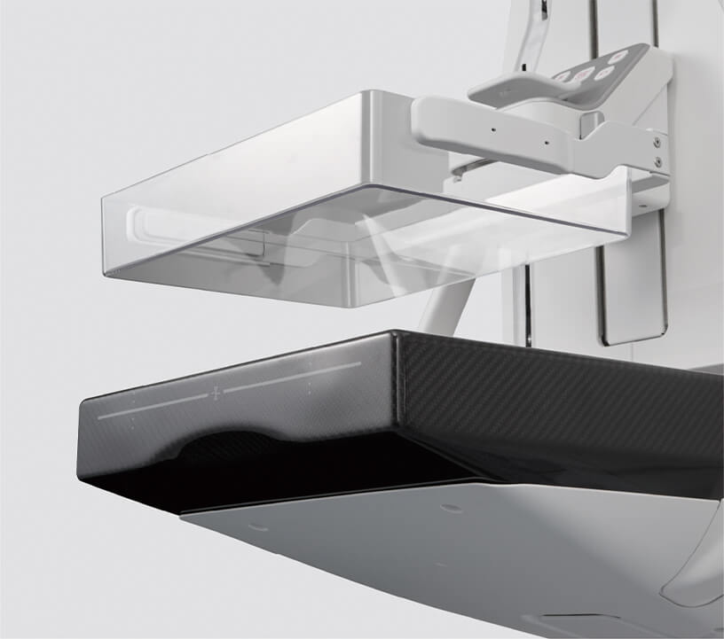
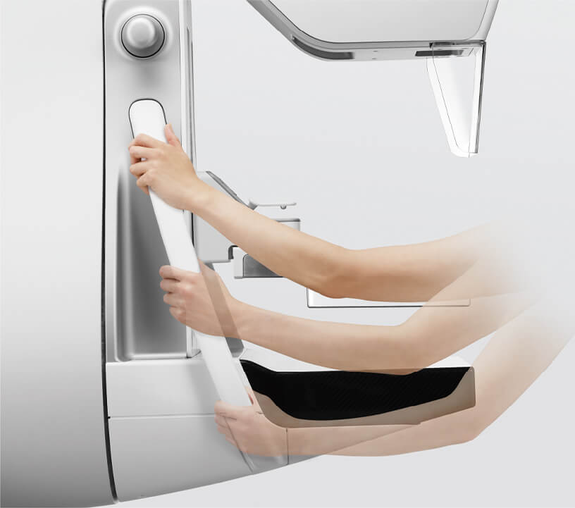
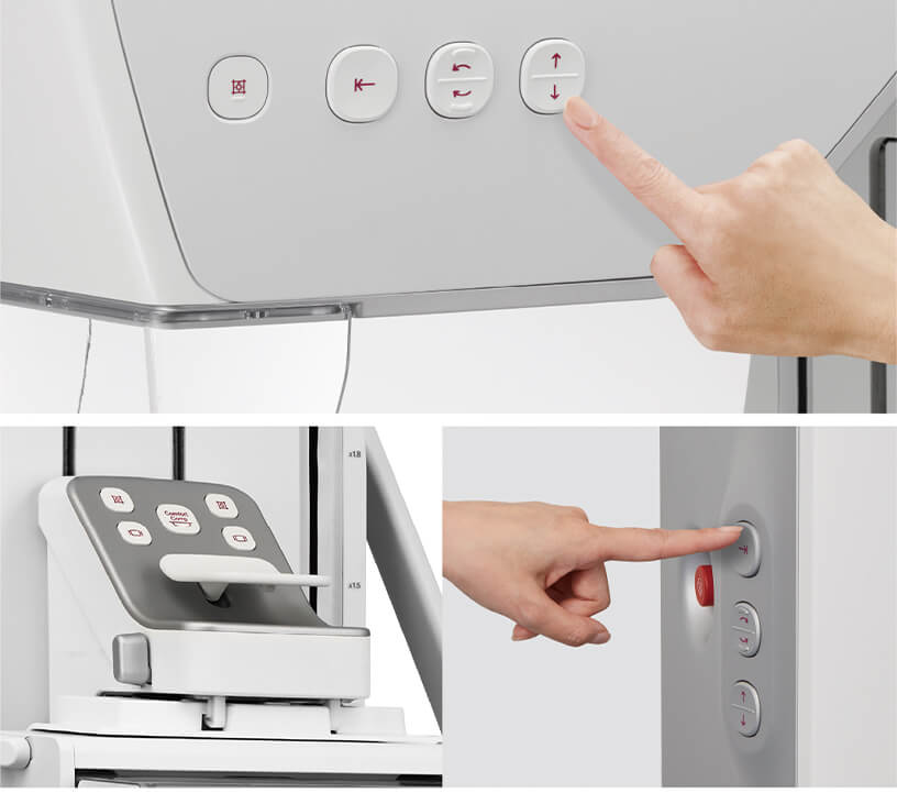
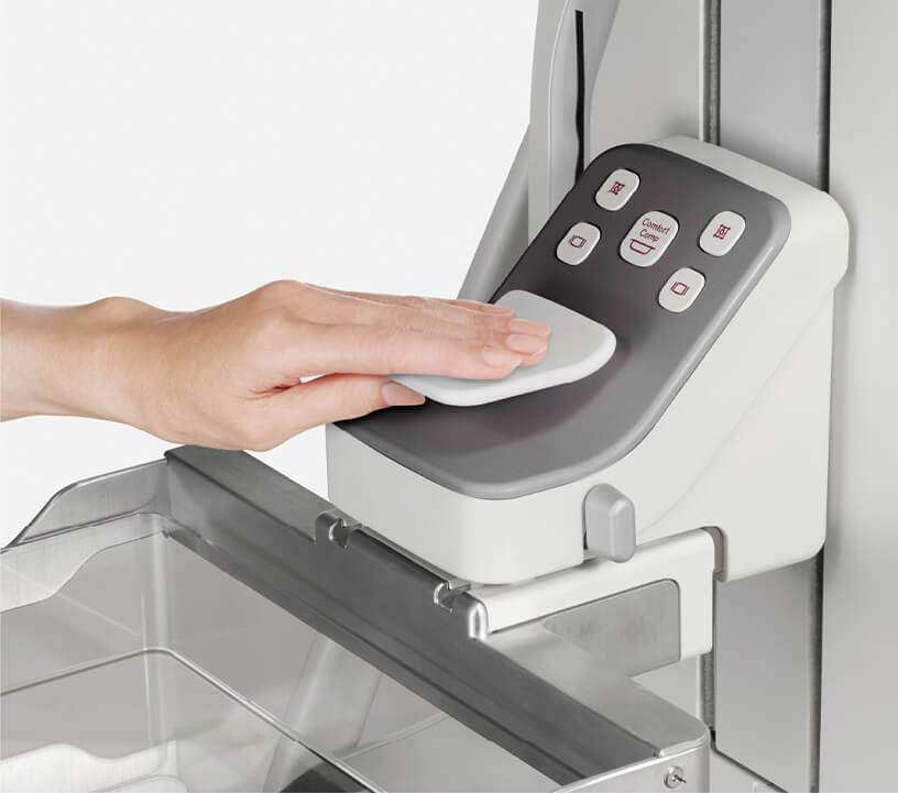
Comfort Comp uses the hysteresis phenomenon to automatically reduce compression pressure within a 3mm range after normal compression, shortening the time at maximum pressure and reducing pain without changing breast thickness or sacrificing image quality. AI-developed positioning support, including the Positioning MAP that projects skin lines and nipple position from past images onto the imaging table, helps technologists capture consistent, comparable images. A thin imaging table with reduced side bulging, a longer armrest, and an accessible button layout make every breast cancer screening exam more comfortable for the patient and easier for staff.
Full Technical Specifications
Complete reference data for capital procurement, RFP submissions, and biomedical review. This specification set is required by breast imaging directors, purchasing teams, and biomedical engineers comparing digital mammography equipment options. Expand each category.
| Detector Type | Direct-conversion flat-panel detector (FPD) |
| Pixel Size | 50 microns, smallest direct-conversion FPD pixel |
| Pixel Arrangement | Hexagonal pattern |
| X-Ray Target | Tungsten target with ISC (Image-based Spectrum Conversion) |
| Image Processing | Dynamic Visualization with MFP, density/contrast adjustment, frequency enhancement, dynamic range compression |
| Standard (ST) Mode | Plus or minus 7.5 degree sweep, 19 shots, 100/150 micron pixels, approx. 5 seconds |
| High Resolution (HR) Mode | Plus or minus 20 degree sweep, 35 shots, 50/100 micron pixels, approx. 12 seconds |
| S-View | Synthesized 2D from tomosynthesis at 50 microns, with breast thickness information (optional) |
| Face Guard T Comfort | Fixed face guard for tomosynthesis, collapses with tube movement at ST and HR angles (optional) |
| Intelligent AEC | Adjusts X-ray dose according to breast type from a low-dose pre-shot image |
| Automatic Sensor Method | Analyzes breast structure, fat density, and implants to select the appropriate sensor |
| Manual Sensor Method | Technician selects mammary gland area during positioning |
| Implant Imaging | Automatically identifies implant area for correct technique |
| Comfort Comp | Automatic decompression within 3mm range after compression, using hysteresis to reduce pain |
| Imaging Table | Thin, compact, reduced side bulging, curved detector front |
| Armrest | Lengthened for comfortable CC and MLO postures, doubles as hand rest |
| AI Positioning MAP | Projects skin lines and nipple position from past images onto the table (optional) |
| Positioning Analysis | AI deep-learning analysis of positioning to improve technique (optional) |
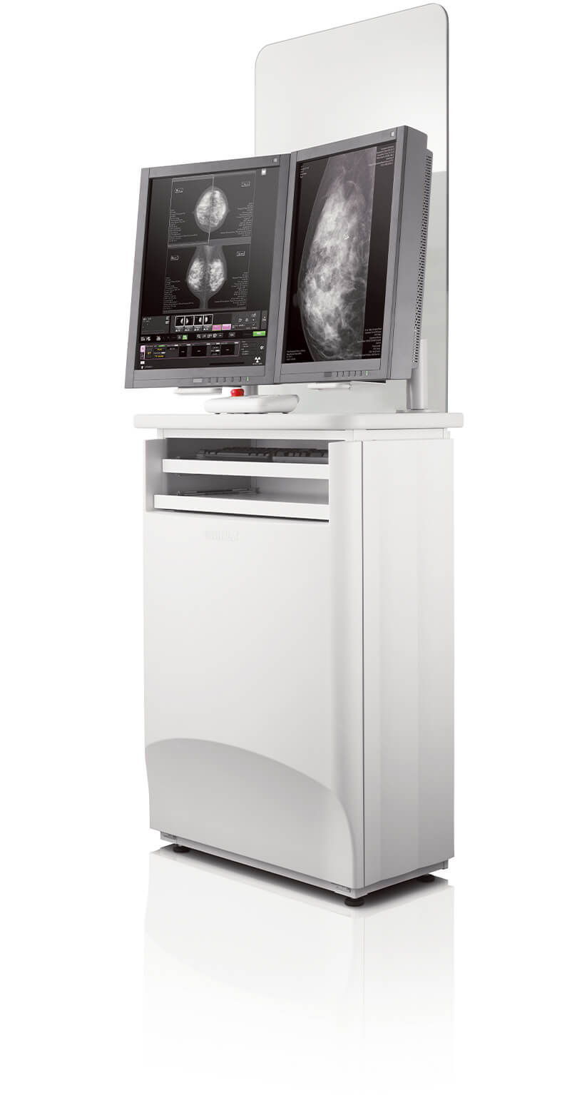
| Acquisition Workstation (AWS) | User-friendly interface with tall, bright screens and integrated X-ray control section |
| Second Monitor | High-definition 3M/5M for current and past image comparison when connected to PACS (optional) |
| Breast Density Assessment | Automatic mammary gland volume analysis output to a DICOM Tag with configurable Density Category (optional) |
| FUJIFILM Mammo QC | 10-item image quality evaluation in 5 minutes with trend graphs and CSV history (optional) |
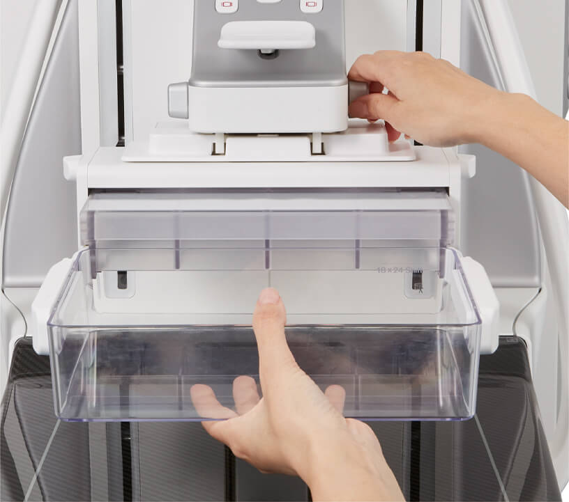
| Shift Compression Plate | 18 x 24 plate moves to center for CC view and top for MLO, with radiation field auto-tracking (optional) |
Clinical Applications
Deployed across the full spectrum of breast cancer screening and diagnostic mammography, wherever image quality, patient comfort, and workflow efficiency drive screening compliance and diagnostic confidence.
Standard tomosynthesis mode with deep depth of field and fast 5-second acquisition supports high-volume breast cancer screening, while Comfort Comp reduces the discomfort that deters patients from returning.
High Resolution tomosynthesis mode with a wider sweep angle and shallow depth of field focuses on areas of interest for close examination, additional imaging, and morphology assessment.
Digital breast tomosynthesis reconstructs focused tomographic slices that reveal lesions hidden by overlapping mammary gland structures, and a single session also produces an S-View synthesized 2D image.
CEDM performs low-energy and high-energy acquisition in one compression and generates an energy subtraction image, providing functional information when an iodinated contrast medium is used.
Intelligent AEC analyzes breast structure, fat density, and implants to set the correct technique automatically, and the Breast Density Assessment function outputs mammary gland volume to a DICOM Tag.
The second monitor displays stereotactic and tomosynthesis biopsy targeting images, supporting image-guided breast biopsy procedures directly from the acquisition workstation.
Acquisition, AI Positioning & PACS Integration
A complete breast imaging solution designed to streamline every step from patient positioning through image acquisition to PACS comparison. The acquisition workstation, AI positioning support, and FUJIFILM Mammo QC keep the mammography workflow efficient and reproducible. Expanded details are in the expandable sections below.
The optimized acquisition workstation (AWS) features a user-friendly interface with tall, bright screens and an integrated X-ray control section, so imaging conditions can be set and confirmed on the same screen. The test screen supports 1, 2, or 4 divided displays, left and right image alignment, and arbitrary image output even during testing. A high-definition 3M or 5M second monitor displays current and past images for comparison when connected to PACS, and automatically displays tomosynthesis reconstructed images.
The Positioning MAP projection function extracts skin lines and nipple positions from past images and projects them onto the imaging table surface, making it possible to capture images that are easily compared with prior studies. The opposite breast can be inverted and projected for left to right comparison. Positioning Analysis uses AI deep-learning to analyze positioning from captured images and help technicians improve their skills, part of Fujifilm’s REiLI medical AI technology brand.
Comfort Comp automatically reduces compression pressure within a 3mm range after normal compression, using the hysteresis phenomenon to shorten the time at maximum pressure. The compression plate can be finely adjusted electrically without removing the line of sight from the breast, and an array of compression paddles sized for each breast and view attach and remove quickly. Arm rotation and height buttons on the tube head enable smoother testing.
FUJIFILM Mammo QC is a quality control program designed exclusively for the Fujifilm Digital Mammography System. A 10-item image quality evaluation completes in 5 minutes, graphically represents day-to-day fluctuations, and manages history in a CSV file. The Breast Density Assessment function automatically analyzes mammary gland volume immediately after imaging and outputs the values to a DICOM Tag with configurable Density Category thresholds.
Regulatory Compliance & Safety
Manufactured by FUJIFILM Corporation, a leader in breast imaging technology and a supporter of the Pink Ribbon Campaign for early detection of breast cancer. Medical Outfitters is an ISO 13485:2016-certified supplier of digital mammography equipment, meaning every Amulet Sophinity ships through a documented, audited quality management system. Full compliance documentation is available in the expandable sections below.
Manufacturer: FUJIFILM Corporation, Tokyo, Japan, a healthcare imaging leader since 1936 and a supporter of the Pink Ribbon Campaign for early breast cancer detection.
Regulatory Approval: All products require the regulatory approval of the importing country. Our team confirms applicable clearances for your region before shipment.
ISO 13485: Medical Outfitters Inc. is independently ISO 13485:2016 certified as a digital mammography equipment supplier.
FUJIFILM Mammo QC: A dedicated quality control program performing a 10-item image quality evaluation in 5 minutes with phantom imaging, visual result entry, and automatic calculation.
Breast Density Reporting: Density Category output to a DICOM Tag supports breast density notification requirements and clinical decision tools.
RoHS: Restriction of Hazardous Substances compliant.
REACH: EU REACH chemical regulation compliant.
Site Planning & Facility Review
The integrated high-voltage design lets the Amulet Sophinity fit in a limited space. Provide your room dimensions, PACS configuration, and the optional packages you need, such as tomosynthesis, S-View, or CEDM, and our team confirms fit and coordinates installation.
Capital Equipment & RFP Support
Volume pricing, mammography equipment cost breakdowns, technical documentation, and logistics coordination for breast imaging program deployments. We support breast imaging directors, biomedical engineers, and hospital purchasing departments evaluating digital mammography equipment and mammography monitors. Contact us for RFP packages.
Frequently Asked Questions
Quick answers to the most common purchase and technical questions about the Fujifilm Amulet Sophinity digital mammography system. Tap any question to expand.
The Fujifilm Amulet Sophinity is a digital mammography system with a 50 micron hexagonal direct-conversion detector, Tungsten target imaging, digital breast tomosynthesis in Standard and High Resolution modes, S-View synthesized 2D imaging, CEDM contrast-enhanced mammography, intelligent AEC, Comfort Comp pain reduction, and AI-based positioning support.
The 3d mammogram cost and overall mammogram price depend on configuration and optional packages such as tomosynthesis, S-View, and CEDM. Diagnostic mammography cost also varies with monitor and workstation selections. Medical Outfitters returns a quote within one business day. Submit a request online or call (305) 885-4045. We also support trade-in evaluations against your current mammography equipment.
Yes. The Amulet Sophinity offers digital breast tomosynthesis with two modes: Standard mode (plus or minus 7.5 degree sweep, 19 shots, approximately 5 seconds) and High Resolution mode (plus or minus 20 degree sweep, 35 shots, approximately 12 seconds, improved depth resolution). An optional S-View synthesized 2D image is generated from the same tomosynthesis acquisition.
The Amulet Sophinity uses the smallest direct-conversion FPD pixel size of 50 microns arranged in a hexagonal pattern, enabling high-definition imaging of microcalcifications at a low radiation dose with a Tungsten target and ISC.
Comfort Comp is an automatic decompression control that reduces compression pressure within a 3mm range after normal compression, using the hysteresis phenomenon to shorten the time at maximum pressure. This reduces patient pain without changing breast thickness or compromising image quality.
Yes. The optional CEDM (Contrast Enhanced Digital Mammography) function performs low-energy and high-energy imaging with a Cu filter in a single compression, then automatically generates an energy subtraction image when an iodinated contrast medium is used.
Yes. By integrating the high-voltage device within the main unit, the overall size is significantly reduced, allowing installation in a limited space. The slim, rounded exterior is also designed to feel less intimidating for patients while remaining aesthetically pleasing for staff.
Through Medical Outfitters Inc., ISO 13485:2016 certified, Miami, FL. Factory-new units with nationwide installation support. Call (305) 885-4045 or request a quote online.
Compatible Equipment
Breast imaging equipment commonly deployed alongside the Fujifilm Amulet Sophinity, from a companion Fujifilm FFDM platform to the specimen radiography and biopsy systems that complete a full breast care suite.

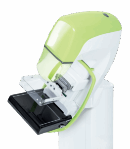
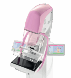
Fujifilm ASPIRE Cristalle
Fujifilm FFDM · HCP detector · DBT · CEDM · high-volume screening
View Product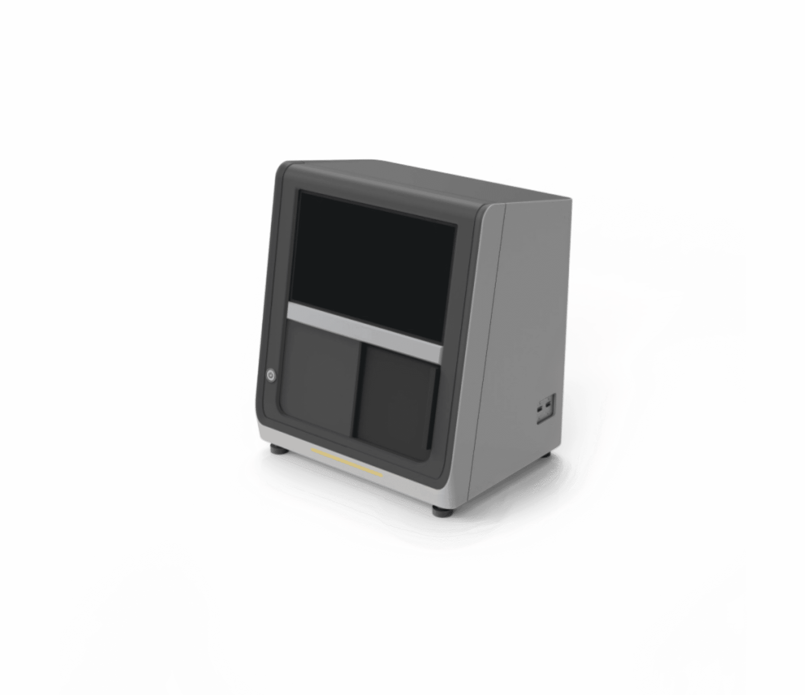
TrueView Core 100Pro Specimen Radiography
Specimen verification · CMOS detector · calcification recognition · PACS/DICOM
View Product
Mammotome Revolve Biopsy System
Vacuum-assisted biopsy · ultrasound-guided · stereotactic · breast biopsy
View ProductRequest a Quote Today
Get in touch with Medical Outfitters today to request your personalized quote and outfit your facility with confidence.