Published 29 September 2026. Written by the Medical Outfitters sales and field engineering team. Frequency ranges and probe designations are typical of current platforms and vary by manufacturer; check the data sheet for the specific probe.
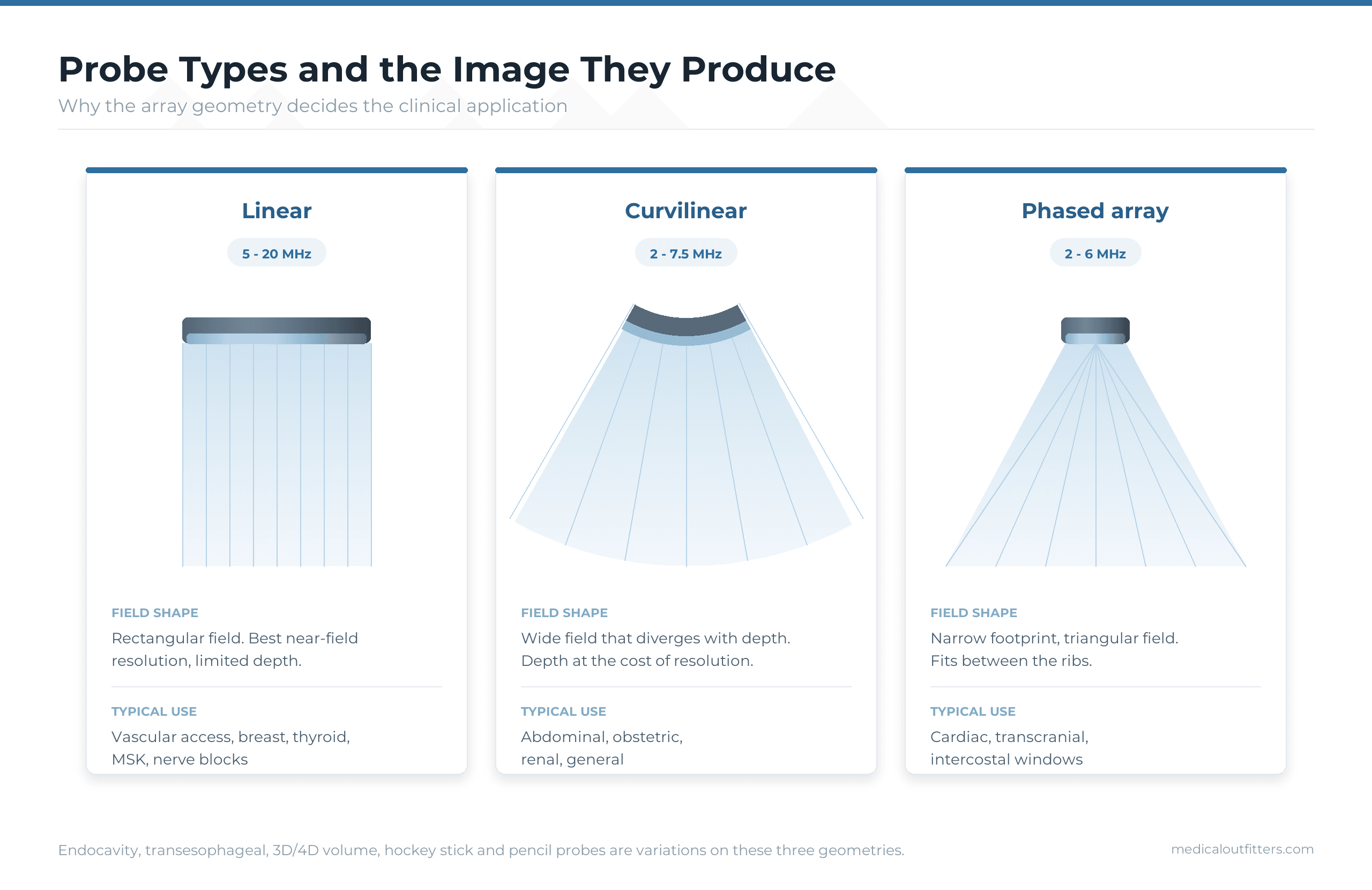
There are three main ultrasound transducer types, linear, curvilinear (convex) and phased array, plus endocavity, transesophageal (TEE), 3D and 4D volume and a handful of specialty probes. The type sets the shape of the image, the frequency sets how deep it can see and how fine the detail is, and the footprint sets where on the body it fits. A linear probe at 5 to 18 MHz images shallow structures in high detail, a curvilinear probe at 1 to 6 MHz images the abdomen and a pregnancy, and a phased array at 1 to 5 MHz images the heart between the ribs.
This explainer goes through every transducer type with its frequency range, footprint, field of view, typical element count and the exams it is bought for, how to read a probe designation such as L12-4 or C5-1, why frequency and depth trade against each other, and why probes are not interchangeable between manufacturers. If you already know the types and want to pick one for a caseload, read how to choose an ultrasound probe; if a probe is faulty, the probe faults, testing and repair guide covers the air scan and the repair-or-replace decision.
- In this guide: What a transducer is
- How transducers are named: L12-4, C5-1, P4-2
- All transducer types compared
- Linear transducers
- Curvilinear (convex) transducers
- Phased array transducers
- Endocavity transducers
- Transesophageal (TEE) transducers
- 3D and 4D volume transducers
- Specialty transducers
- Frequency versus depth
- Compatibility between systems
- Which types a department needs
- Frequently asked questions
What Is an Ultrasound Transducer?
An ultrasound transducer, or probe, converts electrical pulses into sound and the returning echoes back into electrical signals. Inside the head sits an array of piezoelectric elements, usually 128 to 256 of them on a standard probe and thousands on a matrix probe, behind a matching layer and an acoustic lens that focuses the beam. The console fires the elements in sequence, listens for the echoes and builds the image line by line.
The arrangement of those elements is what defines the transducer type: in a straight line, on a curve, or packed into a small block that is steered electronically. Everything else about a probe, its frequency, its footprint and its cable, follows from the exam it was designed for. Our guide to ultrasound probe types, faults and repair covers the acoustic stack in detail and what happens when it fails.
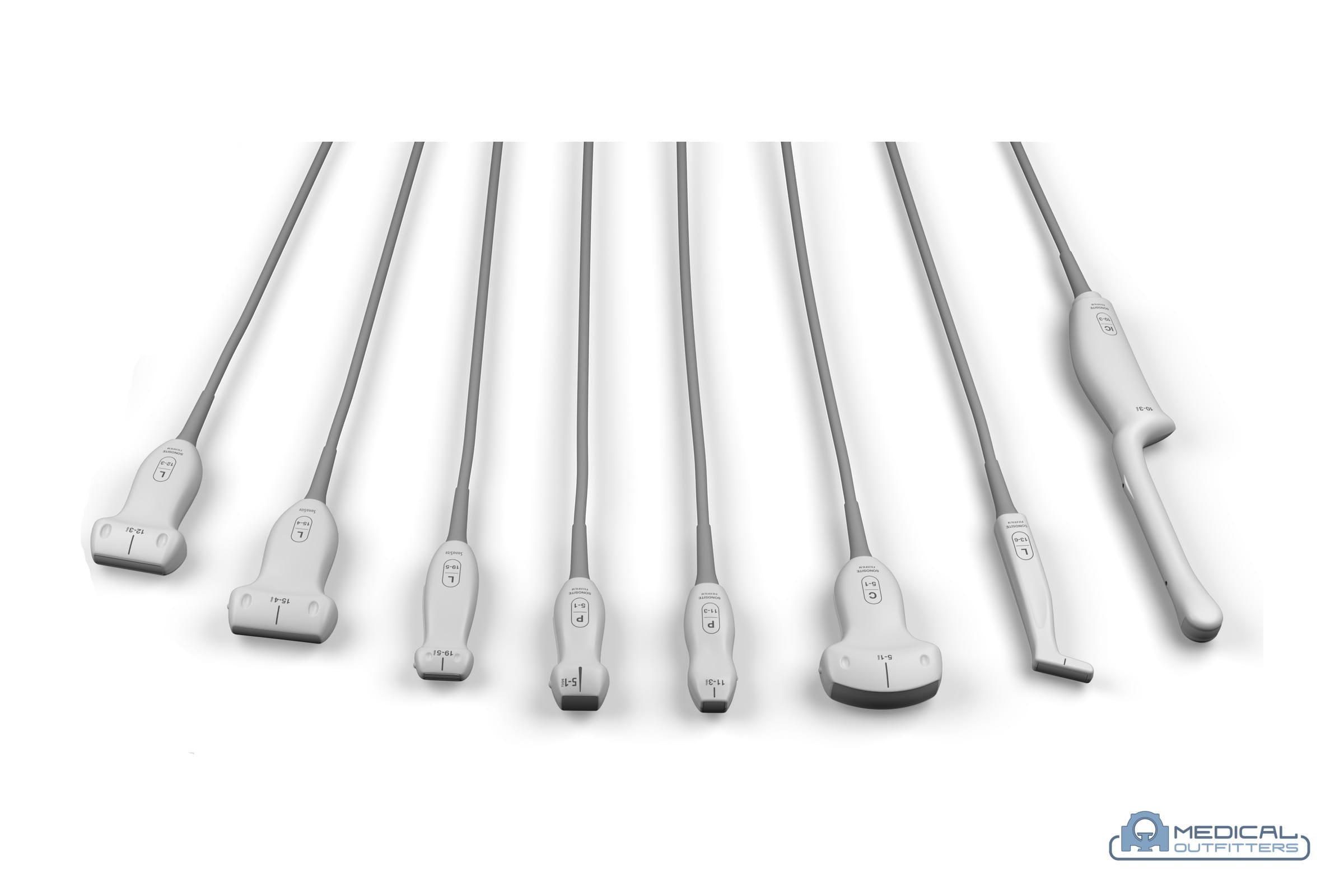
How Are Ultrasound Transducers Named?
Most manufacturers name a probe by the array type and its frequency range, so the designation tells you most of what you need before you read the data sheet.
| Designation | Array type | Frequency range | Example use |
|---|---|---|---|
| L12-4, L18-5, L15-4 | L = linear | 4 to 12 MHz, 5 to 18 MHz, 4 to 15 MHz | Vascular, musculoskeletal, small parts, nerve blocks |
| C5-1, C1-6, C6-2 | C = curvilinear (convex) | 1 to 5 MHz, 1 to 6 MHz, 2 to 6 MHz | Abdomen, obstetrics, general |
| P4-2, P5-1, S4-2 | P or S = phased array (sector) | 2 to 4 MHz, 1 to 5 MHz | Cardiac, transcranial, FAST |
| IC5-9, IC10-3, EC9-4 | IC or EC = intracavity or endocavity | 5 to 9 MHz, 3 to 10 MHz | Transvaginal, transrectal |
| X5-1, X7-2t, 6VT-D | X = matrix, t or T = transesophageal | 1 to 5 MHz, 2 to 7 MHz | 3D cardiac, TEE |
| RAB6-D, RIC5-9-D, V6-2 | R or V = volume (3D/4D), IC = intracavity volume | 2 to 9 MHz | 3D and 4D obstetrics, gynaecology |
| HL or hockey stick L8-18i | Small-footprint linear | 8 to 18 MHz | Superficial musculoskeletal, paediatric, intraoperative |
The two numbers are the bandwidth, from the lowest to the highest frequency the probe can be driven at; the console selects the operating frequency inside that range for each preset. GE, Philips, Siemens, Canon, Mindray, Samsung and Fujifilm each use their own prefixes, so two probes with similar numbers from different manufacturers are similar in role but never physically interchangeable.
All Ultrasound Transducer Types Compared
| Type | Typical frequency | Footprint | Image shape | Depth | Typical exams |
|---|---|---|---|---|---|
| Linear | 5 to 18 MHz (to 22 MHz for superficial work) | Wide, flat face, 25 to 60 mm | Rectangular | 1 to 9 cm | Vascular, MSK, breast, thyroid, small parts, nerve blocks, line placement |
| Curvilinear (convex) | 1 to 6 MHz | Wide, curved face, 50 to 70 mm | Wide sector, fan shaped | Up to 30 cm | Abdomen, obstetrics, pelvis, renal, bladder, general |
| Micro-convex | 3 to 10 MHz | Small curved face, 10 to 20 mm | Tight sector | Up to 20 cm | Paediatric, neonatal, veterinary, intercostal abdominal |
| Phased array (sector) | 1 to 5 MHz | Small square face, about 20 mm | Narrow sector from a point | Up to 30 cm | Cardiac (echo), transcranial, FAST, deep abdominal between ribs |
| Endocavity | 4 to 10 MHz | Small curved face on a long handle | Sector, up to 180 degrees | Up to 15 cm | Transvaginal, transrectal, prostate, early pregnancy |
| Transesophageal (TEE) | 3 to 8 MHz | Small phased array on a steerable insertion tube | Sector, multiplane | Up to 15 cm | Cardiac imaging from the oesophagus in surgery and cardiology |
| 3D and 4D volume | 2 to 9 MHz | Curved or endocavity face with a mechanical sweep, or a matrix array | Volume | Up to 25 cm | Obstetrics, gynaecology, 3D cardiac |
| Pencil (CW Doppler) | 2 to 8 MHz | Tiny, no imaging | None, spectral trace only | Deep | Continuous wave Doppler in cardiac and vascular labs |
Linear Transducers
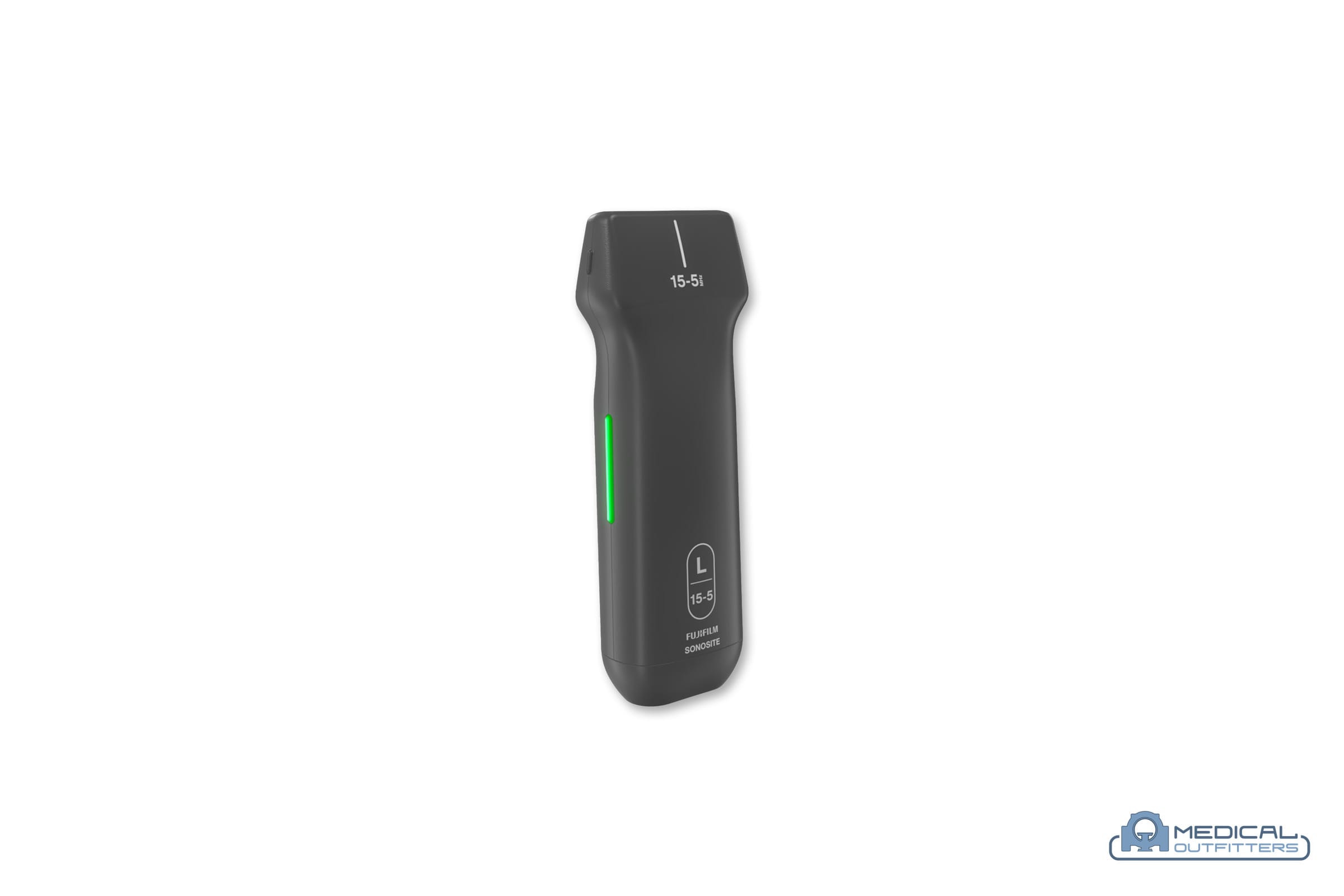
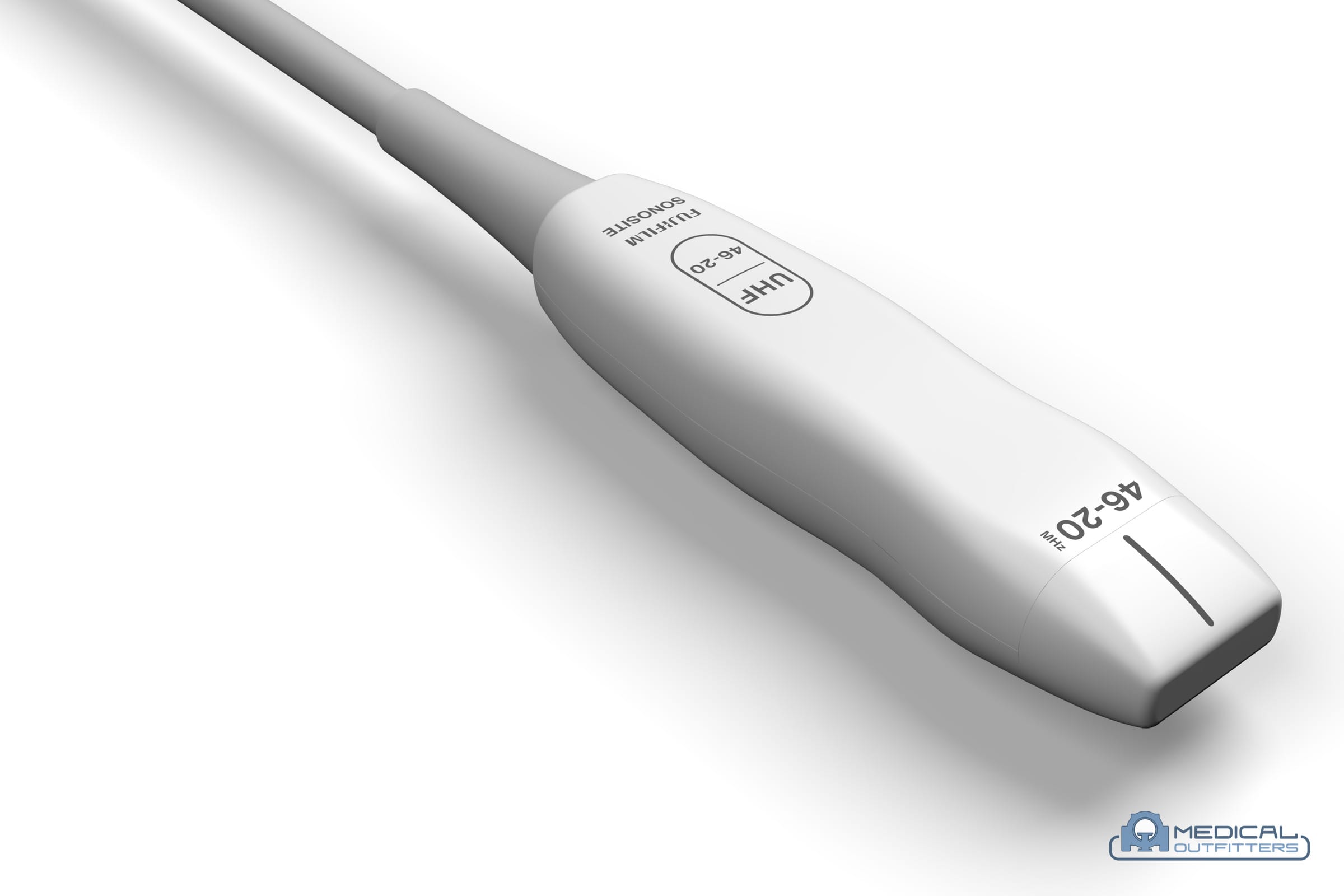
A linear transducer has its elements in a straight row, fires them in groups straight down and produces a rectangular image the same width as the probe face. Because it runs at high frequency, 5 to 18 MHz on general probes and higher on dedicated superficial probes, it gives the finest detail of any type, at the cost of depth: most linear probes image usefully to 6 to 9 cm and the highest frequency ones to 3 or 4 cm.
- Vascular Carotid, venous mapping, DVT studies, arterial Doppler and access for lines
- Musculoskeletal and sports medicine Tendons, ligaments, muscle and joints, with injections under guidance
- Small parts Thyroid, breast, testes, lymph nodes
- Anaesthesia and emergency Nerve blocks and vascular access, where needle visualisation software matters
- Hockey stick and intraoperative linear A short, angled linear array for hands, feet, paediatric and surgical fields
Linear probes are the most handled probes in most departments and the most often damaged: a cracked lens or a split strain relief on a linear probe is the classic repair.
Curvilinear (Convex) Transducers
A curvilinear, or convex, transducer arranges the elements on a curve, so the beams fan out and the image widens with depth. It runs at low frequency, 1 to 6 MHz, which is what lets it reach 25 to 30 cm into the abdomen or a pregnant uterus, and its wide face gives a broad field of view for survey scanning.
- Abdominal Liver, gallbladder, kidneys, spleen, aorta and bladder
- Obstetrics and gynaecology Transabdominal pregnancy scanning and pelvic surveys
- General and emergency The default first probe on most carts and portables
- Micro-convex The same geometry on a small face for neonatal, paediatric, veterinary and intercostal work
The trade-off is resolution: at 2 to 5 MHz the detail is coarser than a linear probe, which is why a curvilinear probe is the wrong tool for a thyroid or a tendon.
Phased Array Transducers
A phased array transducer packs a small block of elements into a footprint about the size of a thumbnail and steers the beam electronically, sweeping it through a sector from a single point. That is what lets it image the heart between the ribs and the brain through the temporal bone window. It runs at 1 to 5 MHz and produces a narrow, triangular image with poorer near-field detail than a linear probe.
- Cardiac Transthoracic echocardiography, the reason most phased array probes are bought
- Transcranial Doppler Through the temporal bone in neurology and critical care
- FAST and point of care A phased array covers heart, lungs and abdomen from one probe in an emergency protocol
- Deep abdominal Where a curvilinear face does not fit between the ribs
Cardiac probes are quoted with the cardiac package and the full Doppler suite, because a phased array without pulsed and continuous wave Doppler cannot do the measurements an echo lab bills for.
Endocavity Transducers
Endocavity transducers, also called intracavity, transvaginal or transrectal probes, put a small curved array on the end of a long handle so it can image from inside the body, where the target is centimetres from the face rather than tens of centimetres. At 4 to 10 MHz they combine the detail of a higher frequency with the short path to the organ.
- Transvaginal Early pregnancy, gynaecology and fertility work
- Transrectal Prostate imaging and biopsy guidance, sometimes with a biplane array
- 3D endocavity Volume versions for gynaecology and prostate
Endocavity probes go through high-level disinfection between every patient, and the disinfection cycle is what shortens their life: fluid ingress at the handle seal and cracked lenses are the common faults, which is why a leakage test belongs in every service visit.
Transesophageal (TEE) Transducers
A transesophageal transducer places a small phased array on a steerable insertion tube, like an endoscope, so the heart can be imaged from the oesophagus with nothing in the way. Adult TEE probes run at 3 to 8 MHz with a multiplane array that rotates through 180 degrees; paediatric and 3D matrix TEE probes are variants. They are used in cardiac surgery, structural heart procedures, intensive care and cardiology when a transthoracic window is poor.
TEE probes are the most expensive probes on most platforms and the most delicate: bite damage to the insertion tube, articulation failures and leak-test failures are the reasons they arrive for repair, and a bite guard and a proper leakage test after every case are the cheapest protection.
3D and 4D Volume Transducers
Volume transducers acquire a block of tissue rather than a slice. Mechanical 3D and 4D probes sweep a curved or endocavity array back and forth inside an oil-filled dome; matrix probes use a two-dimensional array of thousands of elements and steer the volume electronically, which is faster and has no moving parts. 4D simply means 3D in real time.
- Obstetrics Surface rendering of the fetal face and volume assessment, the most common use
- Gynaecology Uterine and endocavity volume imaging
- Cardiac Matrix probes for 3D echo and 3D TEE
Mechanical volume probes have their own failure modes: fluid leaks, bubbles in the dome and worn drive mechanisms, all of which are repairable at a specialist depot, and all of which a saved image and a quick dome inspection will reveal before purchase.
Specialty Ultrasound Transducers
| Transducer | What it is for |
|---|---|
| Pencil (CW Doppler) | A non-imaging continuous wave Doppler probe for high-velocity flow in cardiac and vascular labs |
| Intraoperative and laparoscopic | Small, sterilisable linear or curved arrays for liver, vascular and neurosurgical fields |
| Biplane | Two arrays at right angles in one endocavity or intraoperative head, for prostate biopsy and surgical guidance |
| Intravascular (IVUS) and intracardiac (ICE) | Catheter-mounted arrays used inside vessels and the heart in the cath lab |
| Wireless and handheld | Linear, curved or dual-head probes that carry the whole beamformer and connect to a tablet; a system class as much as a probe type |
| Veterinary | Micro-convex, linear and rectal probes chosen by species; the equipment is the same as human use |
Frequency Versus Depth: The Trade-Off Behind Every Probe
Frequency is the trade that every probe choice comes down to. Higher frequency means shorter wavelength and finer axial resolution, but sound is attenuated faster in tissue, so the beam does not penetrate as far. Lower frequency penetrates deeper at the cost of detail. The console picks an operating frequency inside the probe’s bandwidth for each preset, but it cannot make a linear probe see 20 cm or a curvilinear probe resolve a 1 mm tendon tear.
| Frequency | Practical depth | What it suits |
|---|---|---|
| 1 to 3 MHz | 20 to 30 cm | Deep abdomen, obese patients, cardiac through the ribs |
| 3 to 6 MHz | 10 to 20 cm | General abdominal, obstetric, renal, paediatric |
| 5 to 10 MHz | 5 to 10 cm | Endocavity, vascular, larger MSK structures |
| 10 to 15 MHz | 3 to 6 cm | Thyroid, breast, tendons, nerve blocks, vascular access |
| 15 to 22 MHz | 1 to 3 cm | Superficial MSK, dermatology, paediatric, small animals |
The physics is set out plainly in the NIBIB ultrasound explainer and the RadiologyInfo general ultrasound page; the AIUM official statements cover practice and transducer care.
Are Ultrasound Transducers Interchangeable Between Systems?
Transducers are specific to the platform. The connector, the element pin-out, the console’s beamformer channel count and the software preset for that probe are all matched by the manufacturer, so a GE probe will not plug into a Philips console and a probe designed for a current platform will often not work on the previous generation from the same manufacturer, even when the connector fits. Some probes also need an option code or licence enabled on the console before the system will recognise them.
This is the single fact that costs buyers the most money. Switching manufacturer means replacing the entire probe set, not just the console, and a used console quoted cheaply is only a bargain if the probes you need come with it or exist on the used market. Before buying any probe, confirm the exact console model and software version it will run on, and test it on that port. The ultrasound machine cost guide explains how probe sets move the price of a system.
Which Transducer Types Does a Department Need?
| Department | Core transducer set |
|---|---|
| General and abdominal imaging | Curvilinear plus linear |
| Obstetrics and gynaecology | Curvilinear, endocavity, often a 3D/4D volume probe |
| Cardiology and echo | Phased array, pencil CW Doppler, TEE for surgery and structural work |
| Vascular lab | Linear, plus a curvilinear or phased array for abdominal vessels |
| Musculoskeletal and sports medicine | High-frequency linear and a hockey stick |
| Emergency and critical care | Curvilinear, linear and phased array, or a triple-head handheld |
| Anaesthesia and pain | High-frequency linear with needle visualisation, sometimes a curvilinear for deep blocks |
| Urology | Endocavity or biplane transrectal, curvilinear for renal and bladder |
| Paediatrics and neonatology | Micro-convex, high-frequency linear, small phased array |
| Veterinary | Micro-convex and linear, rectal for large animals |
The next step is the decision itself: which of these for your caseload, at what frequency, on which system. That is the how to choose an ultrasound probe guide. If you are pricing a system with its probes, the ultrasound cost guide has the ranges by class, model and manufacturer, and our ultrasound service page covers probe testing and repair.
Ultrasound Transducer Types: Common Questions
- What are the three main types of ultrasound transducers?
- Linear, curvilinear (convex) and phased array. Linear probes run at high frequency for shallow, detailed imaging of vessels, tendons and small parts; curvilinear probes run at low frequency for deep abdominal and obstetric imaging; phased array probes steer a narrow beam from a small footprint for cardiac imaging between the ribs. Endocavity, transesophageal, 3D/4D volume and specialty probes complete the set.
- What is the difference between a linear and a curvilinear probe?
- A linear probe has a flat face and a straight row of elements, produces a rectangular image and runs at 5 to 18 MHz, so it images shallow structures in fine detail. A curvilinear probe has a curved face, produces a fan-shaped image and runs at 1 to 6 MHz, so it reaches deep into the abdomen at coarser resolution.
- What is a phased array transducer used for?
- Cardiac imaging first of all, because its small footprint fits between the ribs and its electronically steered sector reaches the whole heart. It is also used for transcranial Doppler, FAST exams in emergency medicine and deep abdominal imaging where a curvilinear face does not fit.
- What does L12-4 or C5-1 mean on an ultrasound probe?
- The letter is the array type, L for linear, C for curvilinear, P or S for phased array, IC or EC for endocavity, X for matrix, and the numbers are the bandwidth in megahertz. An L12-4 is a linear probe that can be driven between 4 and 12 MHz; a C5-1 is a curvilinear probe between 1 and 5 MHz.
- Can I use an ultrasound probe from another manufacturer?
- No. Transducers are specific to the platform: the connector, the element pin-out, the beamformer channel count and the software presets are matched by the manufacturer, and some probes need a licence enabled on the console. Switching manufacturer means replacing the whole probe set, which is why probe compatibility should be confirmed before any used console is bought.
- What frequency probe is used for abdominal ultrasound?
- A curvilinear probe in the 1 to 6 MHz range, operated around 2 to 5 MHz depending on the patient. Lower frequencies penetrate deeper for larger patients; higher frequencies give better detail on slim patients and children, where a micro-convex probe at 3 to 10 MHz may be used.
- What is a TEE probe?
- A transesophageal echocardiography probe: a small phased array on a steerable insertion tube that images the heart from the oesophagus, used in cardiac surgery, structural heart procedures and intensive care when the transthoracic window is poor. TEE probes are the most expensive and most delicate probes on most platforms.
- How many transducers does an ultrasound machine need?
- As many as the caseload needs, named in the quote with their frequency ranges. A general clinic usually needs a curvilinear and a linear probe; obstetrics adds an endocavity probe and often a volume probe; cardiology needs a phased array with the cardiac package; emergency departments typically run all three main types.
Tell us what you scan and Medical Outfitters will quote the transducers as part of the system, new or refurbished, with delivery, training and a service plan. We test probes and arrange repair through our service team.