Published 29 September 2026. Written by the Medical Outfitters sales and field engineering team. Frequency ranges and probe designations are typical of current platforms and vary by manufacturer; check the data sheet for the specific probe.
Choose an ultrasound probe by working through five questions in order: what you need to image, how deep it is, what window you have to image through, which system it must run on, and what the probe has to survive. The exam decides the array type, the depth decides the frequency, the window decides the footprint, the system decides which probes are even eligible, and the workload decides whether new, refurbished or repaired is the right buy.
This guide turns those five questions into a decision, exam by exam, with the probe families that fit each caseload, the frequency-to-depth rule, the compatibility checks that stop expensive mistakes, and how to buy a probe without paying for one you do not need. If you want the reference on what each type is, start with ultrasound transducer types explained; if the question is whether a failing probe is worth fixing, the probe faults, testing and repair guide covers that.
- In this guide: Step 1: match the array type to the exam
- Step 2: choose the frequency for the depth
- Step 3: fit the footprint to the window
- Step 4: confirm the probe runs on your system
- Step 5: features that change what a probe can do
- Step 6: new, refurbished or repaired
- How many probes a department needs
- Mistakes that cost money
- Frequently asked questions
Match the Array Type to the Exam
Start with the exam, because the array type follows from it. The table gives the probe family that does most of the work in each area and the second probe that usually joins it.
| Exam or specialty | First probe | Second probe | Why |
|---|---|---|---|
| Abdomen, liver, kidneys, bladder | Curvilinear C1-6 or C5-1 | Phased array for intercostal views | Depth and a wide survey field |
| Obstetrics | Curvilinear C1-6 | Endocavity IC5-9 for early pregnancy; 3D/4D volume probe | Transabdominal depth plus early and volume work |
| Gynaecology and fertility | Endocavity IC5-9 or EC9-4 | Curvilinear | Detail from inside the pelvis |
| Cardiology and echo | Phased array P4-2 or P5-1 | Pencil CW Doppler; TEE for surgery and structural work | Fits between the ribs, steers the whole heart |
| Vascular | Linear L12-4 or L9-3 | Curvilinear for abdominal vessels | High detail on vessel walls and flow |
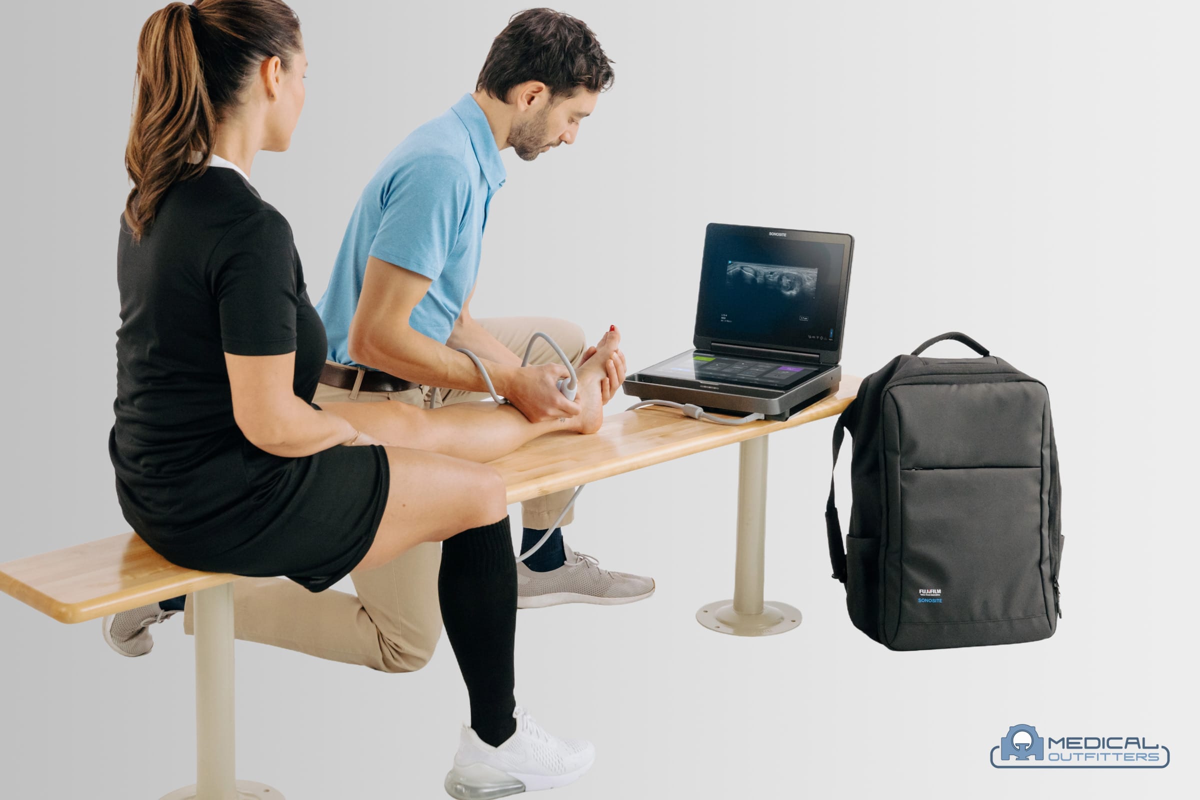
| Musculoskeletal and sports medicine | Linear L18-5 or L15-4 | Hockey stick L8-18 for hands, feet and superficial tendons | Fine detail at 1 to 6 cm |
| Breast, thyroid, small parts | Linear L12-4 to L15-4 | None usually needed | High resolution, shallow |
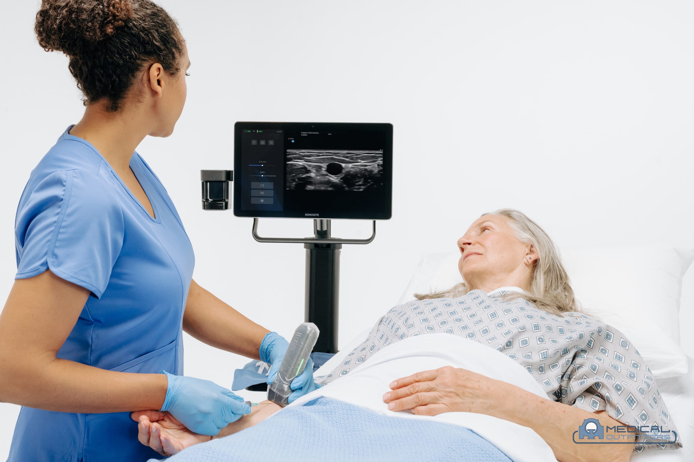
| Anaesthesia and nerve blocks | Linear L12-4 with needle visualisation | Curvilinear for deep blocks and spine | Needle guidance in the near field |
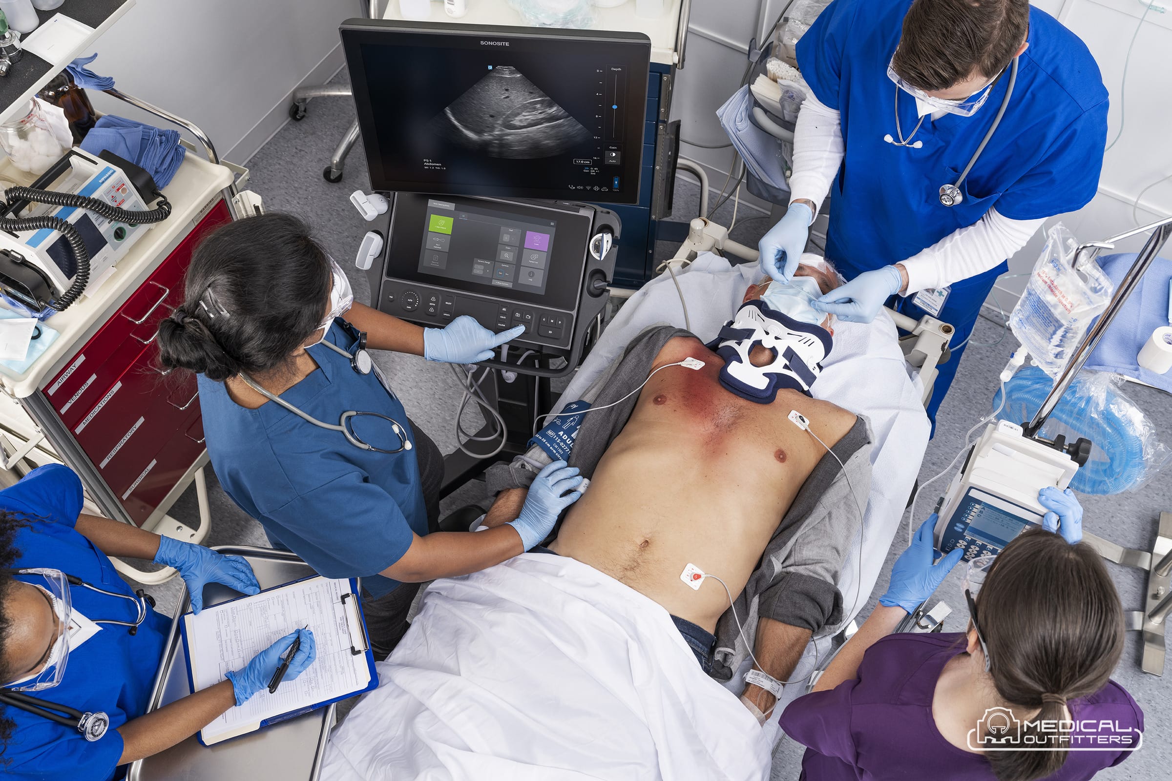
| Emergency and critical care | Curvilinear, linear and phased array | Or a triple-head handheld | One cart covers FAST, lungs, lines and abdomen |
| Urology and prostate | Endocavity or biplane transrectal | Curvilinear for renal and bladder | Biopsy guidance |
| Paediatrics and neonatology | Micro-convex C8-5 and high-frequency linear | Small phased array for hearts | Small footprints, shallow targets |
| Veterinary | Micro-convex and linear | Rectal probe for large animals | Species and size set the frequency |
Choose the Frequency for the Depth
Every probe is a bandwidth, and the rule is simple: the deeper the target, the lower the frequency you need, and the lower the frequency, the coarser the detail. Pick the highest frequency that still reaches the deepest structure you scan on your typical patient, then make sure the probe’s range covers the larger patients too.
| Target depth | Frequency to look for | Probe family |
|---|---|---|
| 1 to 3 cm: skin, superficial tendons, small animals | 15 to 22 MHz | High-frequency linear, hockey stick |
| 3 to 6 cm: thyroid, breast, vascular access, nerve blocks | 10 to 15 MHz | Linear L12-4, L15-4 |
| 5 to 10 cm: larger MSK, vascular, endocavity | 5 to 10 MHz | Linear L9-3, endocavity IC5-9 |
| 10 to 20 cm: abdomen, obstetrics, paediatric | 3 to 6 MHz | Curvilinear C5-1, micro-convex |
| 20 to 30 cm: deep abdomen, bariatric, cardiac | 1 to 3 MHz | Curvilinear C1-6, phased array P4-2 |
Broadband probes soften the choice, because a C1-6 covers both a slim patient at 5 MHz and a bariatric patient at 1.5 MHz. What broadband cannot do is turn a linear array into an abdominal probe or a curvilinear array into a thyroid probe: the array geometry still sets the image shape.
Fit the Footprint to the Window
The footprint is the size and shape of the face that touches the patient, and it has to fit the acoustic window, the gap in bone or gas through which the sound can travel. A wide curvilinear face is ideal on an abdomen and useless between ribs; a phased array’s thumbnail-sized face fits between ribs and through the temporal bone; a micro-convex fits a neonate or a small animal; an endocavity probe removes the window problem by imaging from inside.
- Ribs and clavicle Phased array or micro-convex
- Neonatal head Micro-convex or small phased array through the fontanelle
- Pelvis and prostate Endocavity
- Hands, feet, wrists and small joints Hockey stick linear
- Open abdomen and obstetrics Full-size curvilinear
- Surgical field Intraoperative or laparoscopic probes that can be sterilised or bagged
Confirm the Probe Runs on Your System
This is the step that saves the most money, because a probe that does not run on your console is worth nothing to you. Transducers are matched to the platform by connector, element pin-out, beamformer channel count and software preset, and they are not interchangeable between manufacturers or, often, between generations of the same manufacturer.
- Confirm the console model and software version A probe supported on the current release may not appear on an older one
- Confirm the probe is on the compatibility list for that console By exact model designation, not by family
- Check for licences and option codes Some probes, and most advanced modes on them, need an option enabled on the console
- Count the ports A three-port console with four probes means swapping connectors all day, which is how connector pins get bent
- Test it on that port A used probe should be air-scanned and imaged on the console it will live on before you pay for it
Our explainer on ultrasound transducer types covers the naming conventions by manufacturer, and the ultrasound cost guide explains why a console quoted without the right probes is not comparable to one quoted with them.
Features That Change What a Probe Can Do
Two probes of the same type and frequency can differ in what they can do, and the difference is usually a feature or a licence rather than the array.
| Feature | What it adds | Who needs it |
|---|---|---|
| Needle visualisation and needle guides | Enhances the needle in the near field, or clips a mechanical guide to the probe | Anaesthesia, vascular access, biopsy |
| Elastography | Tissue stiffness maps, licensed on the console and supported on specific probes | Liver, breast, thyroid, MSK |
| Contrast-enhanced ultrasound | A licence and a probe that supports contrast-specific imaging | Liver and renal characterisation |
| Matrix and 4D | Real-time volume imaging without a mechanical sweep | Obstetrics, 3D echo |
| Biplane | Two arrays at right angles for simultaneous planes | Prostate biopsy, intraoperative |
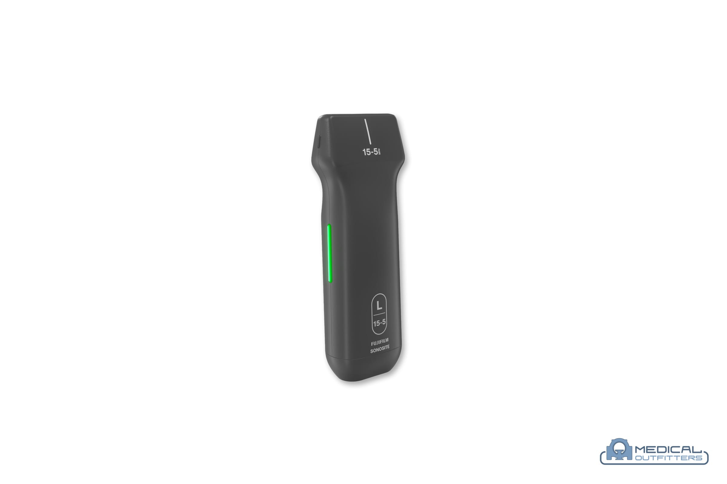
| Wireless and handheld | Beamformer in the probe, image on a tablet | Point of care, home and field programs |
| Ruggedised and sealed | Drop resistance and full immersion for disinfection | Emergency, endocavity, veterinary |
New, Refurbished or Repaired?
Probes carry much of the value of an ultrasound system, so the buy decision on a probe deserves the same care as the console. A new OEM probe runs from a few thousand dollars for a standard linear or curved probe to well into five figures for TEE, matrix and volume probes; a refurbished or exchange probe of the same type is usually 40 to 60 per cent less, and a repair of a damaged probe is normally a fraction of either.
- New When the probe is a current model you will run for years on a supported platform, or when the feature you need (matrix, 4D, contrast) only exists on the current generation
- Refurbished or exchange When the platform is established and the probe type is common; insist on a test report, an air scan and a warranty that names the probe
- Repair When an existing probe has a split strain relief, a damaged cable, bent connector pins, a cracked lens over an intact array or dropout on one or two elements; test first, then decide
- Replace rather than repair When the array has extensive element loss, the probe has fluid ingress, or the platform is leaving support
Test before you spend anything: an air scan at high gain shows element dropout as dark bands, a flex of the cable reveals a crackle, and a second port or a second system separates a probe fault from a console fault. The routine is in our probe faults, testing and repair guide, and we test transducers and arrange probe repair through our service team. Every refurbished ultrasound machine we sell is quoted with tested probes; see where to buy a refurbished ultrasound machine for the checks.
How Many Probes Does a Department Need?
Enough to cover the caseload without swapping connectors all day, and no more, because every probe is an asset that has to be disinfected, tested and eventually repaired.
| Setting | Typical set |
|---|---|
| Single-specialty clinic (vascular, MSK, thyroid) | One or two linear probes, sometimes a hockey stick |
| General practice or urgent care | Curvilinear plus linear |
| Obstetrics and gynaecology practice | Curvilinear, endocavity, volume probe |
| Cardiology office | Phased array, pencil CW; TEE only if the practice does procedures |
| Emergency department | Curvilinear, linear, phased array on each cart, or a triple-head handheld per bay |
| Hospital radiology | Curvilinear, two linear frequencies, phased array, endocavity, micro-convex and often a volume probe |
| Veterinary practice | Micro-convex and linear; rectal for equine and bovine work |
Probe-Buying Mistakes That Cost Money
- Buying by brand instead of by exam The badge does not decide what the probe can image; the array type, frequency and footprint do
- Buying a console without the probes A cheap console with two tired probes costs more than a fairly priced one with five tested probes, because re-probing a system can approach the price of the system
- Assuming Doppler and advanced modes are included Colour, pulsed wave, continuous wave, elastography and contrast are frequently licensed separately; confirm in writing
- Ignoring disinfection An agent that is not on the manufacturer’s approved list damages lenses and voids cover; endocavity and TEE probes need a leakage test after high-level disinfection
- Skipping the test An air scan takes two minutes and is the difference between a probe and a paperweight
- Too few ports Constant connector swapping is how pins get bent and how the most expensive probe in the room ends up on the floor
Choosing an Ultrasound Probe: Common Questions
- How do I choose the right ultrasound probe?
- Work through five questions in order: what you need to image (which sets the array type), how deep it is (which sets the frequency), what window you have to image through (which sets the footprint), which console it must run on (which decides eligibility), and what it has to survive (which decides features and build). Then decide between new, refurbished and repaired based on the platform’s support and the probe’s test results.
- Which ultrasound probe is used for abdominal scanning?
- A curvilinear probe in the 1 to 6 MHz range, such as a C1-6 or C5-1, with a phased array as a second probe for views between the ribs. Slim patients and children may be better served by a micro-convex probe at 3 to 10 MHz.
- Which probe is used for vascular ultrasound?
- A linear probe in the 3 to 12 MHz range for carotid, venous and arterial work, with a higher-frequency linear probe for superficial vessels and vascular access, and a curvilinear or phased array probe for the abdominal aorta and deep vessels.
- What frequency should an ultrasound probe be?
- As high as will still reach the deepest structure you scan. Superficial work at 1 to 3 cm uses 15 to 22 MHz, thyroid, breast and vascular access use 10 to 15 MHz, endocavity and larger MSK use 5 to 10 MHz, abdominal and obstetric imaging uses 3 to 6 MHz, and deep abdominal and cardiac imaging uses 1 to 3 MHz.
- Can I use any ultrasound probe with any machine?
- No. Probes are matched to the platform by connector, element pin-out, beamformer channel count and software presets, and some need a licence enabled on the console. Confirm the exact console model and software version, check the compatibility list by probe designation, and test the probe on that port before paying.
- Is a refurbished ultrasound probe a good buy?
- Yes when the platform is established, the probe type is common and the seller supplies a test report, an air scan and a warranty that names the probe. A refurbished or exchange probe usually costs 40 to 60 per cent less than new. Insist on testing it on the console it will run on.
- Should I repair or replace a damaged ultrasound probe?
- Repair when the fault is a split strain relief, a damaged cable, bent connector pins, a cracked lens over an intact array or dropout on one or two elements. Replace when the array has extensive element loss, the probe has fluid ingress or the platform is leaving support. Test first with an air scan; many probes written off as failed are repairable.
- How many probes should an ultrasound machine come with?
- Enough to cover the caseload: a general clinic needs a curvilinear and a linear, obstetrics adds an endocavity and often a volume probe, cardiology needs a phased array with the cardiac package, emergency departments run all three main types, and hospital radiology usually carries six or more. Quote the probes with the system so you are comparing like with like.
Medical Outfitters quotes transducers as part of the system, new or refurbished, tests every probe before it ships and arranges probe repair through its service team. Serving the US, Puerto Rico and the Caribbean.