What is a C arm, and why does nearly every operating room, surgery center and pain clinic in the country own one? A C-arm is the imaging system that lets a surgeon see inside the patient while the procedure is happening: the fracture being reduced, the screw entering the pedicle, the needle reaching the facet joint, the catheter moving through the vessel. It rolls in on wheels, it needs no shielded room of its own, and it turns any procedure room into an imaging room for as long as the case lasts. Because it is so common, the term gets used loosely, and buyers meet a long list of variations: mini C-arms, full-size C-arms, flat-panel and image intensifier systems, 3D C-arms and fixed C-arms in cath labs. This guide explains what a C-arm is and how it works, how C-arm fluoroscopy differs from a plain X-ray, the types and what each is for, how dose is controlled and regulated, and what a C-arm machine costs to buy or rent in 2026.
Medical Outfitters sells new Ziehm, Orthoscan and Fujifilm C-arms as an authorized dealer and certified refurbished GE and Siemens systems through MO Select, delivered, installed and serviced across the United States, Puerto Rico and the Caribbean, according to our C-arm machine cost guide. We wrote this for the surgery center administrator, the practice owner, the OR manager and the clinician who has to explain to a board what the machine is before anyone approves buying one. If you only need the price, the cost guide has every band by type and model.
- What it is: a mobile fluoroscopy system with the X-ray tube and the detector held on a C-shaped arm, used for live imaging during surgery and procedures.
- How it works: the tube fires short pulses of X-rays through the patient, the detector turns them into video frames, and the monitor shows the anatomy and the instruments in real time.
- The types: mini C-arms for hands and feet, full-size mobile C-arms for the operating room, and 3D C-arms that add a CT-like volume.
- Dose rules: 21 CFR 1020.32 caps the air kerma rate at 88 mGy per minute in normal mode and requires a dose display and last-image-hold on systems made since June 10, 2006.
- Cost: $55,000 to $135,000 refurbished and $165,000 to $295,000 new for a full-size mobile C-arm, delivered, installed and warrantied, per the Medical Outfitters C-arm cost guide.
What Is a C Arm? The Machine Behind the Name
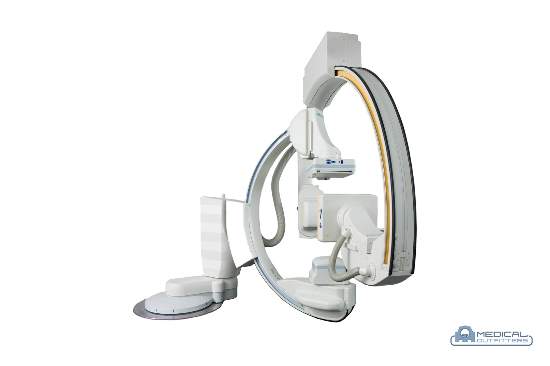
A C-arm is an X-ray system built around a single curved arm shaped like the letter C. The X-ray tube sits at the bottom end of the arm and the image receptor, either an image intensifier or a flat-panel detector, sits at the top. The patient lies on the operating table in the open mouth of the C, between the two. Because the tube and the detector are fixed to the same arm, they always face each other at the correct distance, and the whole arm can slide around its own curve (orbital rotation), tilt sideways (angulation), move up and down, and swing horizontally without the patient being moved at all. That freedom is the whole point: the surgeon gets a front view, a side view or any oblique angle in a few seconds.
What does C arm stand for? Nothing beyond the shape. The term is descriptive, like the U-arm used on some radiography systems; it is not an acronym. People also call it a C arm machine or a C arm X ray system, and say mini C arm for the small extremity version; all of these describe the same family of mobile fluoroscopy equipment, and the Medical Outfitters C-arm vs fluoroscopy explainer sorts out the related terms.
A mobile C-arm is usually two units on wheels. The first is the C-arm stand itself, which carries the generator, the tube, the arm and the detector, and is steered to the table with handles and brakes. The second is the monitor cart or workstation, with one or two displays, the image processing computer, storage, and the connections to the hospital network and PACS. Some compact systems, such as the Ziehm Solo FD and the Fujifilm Persona CS listed in our cost guide, put everything on one chassis for small rooms. The components that matter to a buyer are:
- The generator and tube: from 2.4 kW monoblock units for pain and orthopedic work to 15 to 25 kW rotating-anode systems for vascular and bariatric cases, per the Medical Outfitters C-arm cost guide.
- The detector: a 9 or 12 inch image intensifier on older systems, or a 21 to 31 cm CMOS flat panel on current ones.
- The arc: its depth and its orbital range decide how large a patient fits and how far the arm can rotate around the table.
- The software: image processing, dose management, and licensed options such as vascular subtraction, cardiac imaging or 3D reconstruction.
Fixed C-arms exist too. Cath labs and angiography suites use C-arms mounted on the floor or the ceiling, with larger detectors and far more powerful generators, as part of a shielded room; our guide to what a cath lab is covers them. When people search for a C-arm, however, they almost always mean the mobile kind, and that is the machine this article is about. A fixed radiography and fluoroscopy room, the other fixed fluoroscopy installation, is priced in our fluoroscopy room cost guide.
How C-Arm Fluoroscopy Works: From X-Ray Pulse to Live Image
A conventional C-arm X-ray is not a single still picture. A radiography room fires one short, strong exposure and records one image. A C-arm instead runs fluoroscopy: a stream of low-intensity exposures, each turned into a video frame, so the monitor shows motion in real time. The FDA’s fluoroscopy page lists the uses that rely on it, from guiding catheters and placing stents to orthopedic surgery such as joint replacement and fracture treatment.
Pulsed fluoroscopy. Current C-arms rarely run the beam continuously. They fire short pulses a set number of times a second, and the software fills the gaps between them so the image still looks smooth. Fewer pulses means less radiation, so the operator picks the lowest pulse rate that still shows the motion clearly: a slow pulse rate for checking a screw position, a faster one for following contrast through a vessel.
Automatic dose rate control. As the arm moves from a thin wrist to a thick pelvis, the system adjusts the tube voltage and current automatically to keep the image bright enough to read. Federal rules make that automatic control mandatory on any system that can exceed 44 mGy per minute, under 21 CFR 1020.32(d).
Last-image-hold and saved images. When the operator releases the foot pedal, the last frame stays on the screen, so the surgeon can study it with the beam off. Under 21 CFR 1020.32(j), every fluoroscopic system made on or after June 10, 2006 must have this feature. Frames worth keeping are saved as digital spot images and sent to PACS, which become the intraoperative record. What the PACS that stores them costs is in our PACS system cost guide.
Vascular modes. On higher-power systems, licensed software adds digital subtraction angiography, which removes the bone and soft tissue so only the contrast-filled vessels remain, and roadmapping, which overlays a saved vessel map on the live image to guide a wire. These options are licensed separately and, as our cost guide notes, do not always transfer with a used machine.
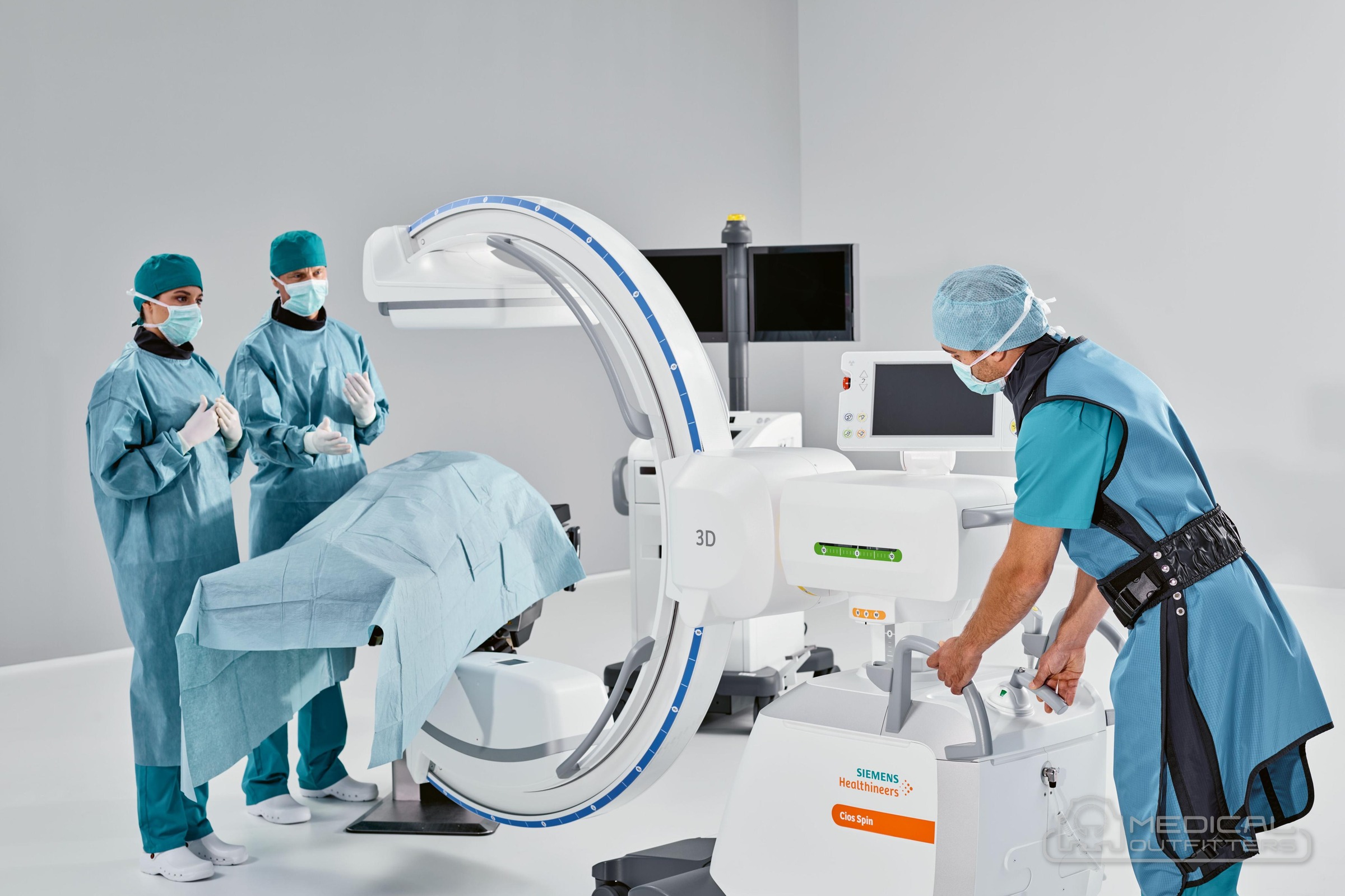
Image Intensifier vs Flat Panel: The Biggest Difference Between C-Arms
Two C-arms can look almost identical from across the room and produce very different images, because of what sits at the top of the arm. The detector is the first thing to check on any C-arm, new or used, and it is the second biggest factor in the price after the format, according to the Medical Outfitters C-arm cost guide.
The image intensifier is the older technology: a large vacuum tube that turns X-rays into light, amplifies the light and passes it to a video camera. It works, it is well understood, and refurbished intensifier systems such as the GE OEC 9800 and 9900 remain common in surgery centers. Its limits are built in. The image is round, it bends slightly at the edges (geometric distortion), its brightness fades as the tube ages, and the bulky housing takes up room above the patient.
The flat-panel detector, usually CMOS on current mobile systems, converts X-rays directly into a digital image on a thin, rectangular plate. The image is square or rectangular with no distortion, has a wider range of gray tones, and generally needs less dose for the same image quality, as the Medical Outfitters C-arm vs fluoroscopy explainer notes. GE HealthCare offers its OEC Elite CFD with a 21 cm or 31 cm CMOS detector and says on its product page that the design shows up to 22 percent more anatomy than monoblock C-arms. Siemens Healthineers lists a 30 x 30 cm CMOS flat detector on the Cios Spin.
| Feature | Image intensifier | Flat-panel detector |
|---|---|---|
| Image shape | Round | Square or rectangular |
| Distortion | Some at the edges | None |
| Aging | Brightness fades, burn-in possible | Dead pixels, needs recalibration |
| Dose for the same image | Higher | Generally lower |
| Typical systems | GE OEC 9800 and 9900, Siemens Arcadis | GE OEC Elite CFD, Siemens Cios, Ziehm Vision FD, Fujifilm Persona C |
| Price position | Lower half of each band | Upper half; holds value longer |
The practical rule from our cost guide: a flat panel C-arm is the better buy for a system you plan to keep for years, but a healthy image intensifier is a better purchase than a tired flat panel. On a used system, ask for the intensifier’s brightness and burn history or the panel’s dead-pixel map and calibration record before you compare prices. The used C-arm buying guide lists the full set of checks.
Types of C-Arm: Mini, Full-Size and 3D
C-arms come in three formats, and the format decides almost everything else: what cases the machine can image, what room it needs and which price band it falls in. Buying the wrong format in either direction is the most expensive mistake in the category, as our cost guide puts it.
Mini C-arm. A mini C-arm is a small, low-power system built for the extremities: hands, wrists, elbows, feet and ankles. The arm is short, the detector is small and the dose is the lowest in the category, so it fits a hand surgery room, a podiatry office or an orthopedic procedure room. The Orthoscan TAU family is the common example, with flat detectors of 15 x 12 cm on the TAU 1512, 15 x 15 cm on the TAU 1515 and 20 x 20 cm on the TAU 2020. Federal rules treat these short systems separately: a C-arm made on or after June 10, 2006 with a source-to-detector distance under 45 cm must be labeled for extremity use only, under 21 CFR 1020.32(g). A mini C-arm cannot image a spine, a hip or a vessel.
Full-size mobile C-arm. This is the operating-room workhorse for orthopedics, trauma, spine, pain management, urology and general surgery, and the machine most buyers mean when they say C-arm. It has a larger arc, a 9 to 12 inch image intensifier or a 21 to 31 cm flat panel, and a generator that ranges from a compact monoblock to a high-power rotating-anode system for vascular and bariatric work. The Ziehm Solo FD, the Ziehm Vision FD, the Fujifilm Persona C, the GE OEC line and the Siemens Cios family all sit here.
3D C-arm. A 3D C-arm rotates its arm around the patient while taking hundreds of images, then reconstructs a cone-beam CT volume in the operating room. Siemens Healthineers lists up to 400 projections, a 30 second scan and a reconstructed volume of 25 x 25 x 16 cm for the Cios Spin. The surgeon can check screw positions in the spine or the alignment of a joint fracture before closing, instead of finding a problem on a postoperative CT. The Ziehm Vision RFD 3D, which our cost guide notes was the first mobile 3D C-arm with a flat-panel detector, is the other main platform.
Hybrids. The Fujifilm FDR Cross combines a C-arm and a portable X-ray in one battery-powered unit, for sites that need both but have room or budget for only one.
What a C-Arm Is Used For in Surgery and Procedures
A C-arm earns its place wherever a clinician needs to see bone, hardware, a needle or contrast while working. The FDA lists orthopedic surgery, catheter guidance, stent placement and angiography among the uses of fluoroscopy, and the Medical Outfitters C-arm guide groups the work into five areas.
- Orthopedics and trauma: fracture reduction, plate and nail placement, joint replacement and hardware checks. This is the largest single use of full-size and mini C-arms.
- Spine: pedicle screw placement, kyphoplasty and fusion, where a 3D C-arm can confirm screw position in the room.
- Pain management: epidural steroid injections, facet and medial branch blocks, radiofrequency ablation and spinal cord stimulator trials, all done with the needle guided under live imaging.
- Urology: kidney stone procedures, ureteral stents and retrograde studies.
- Vascular, cardiac and GI: angiography, catheter and pacemaker lead placement and ERCP on the higher-power systems with vascular software.
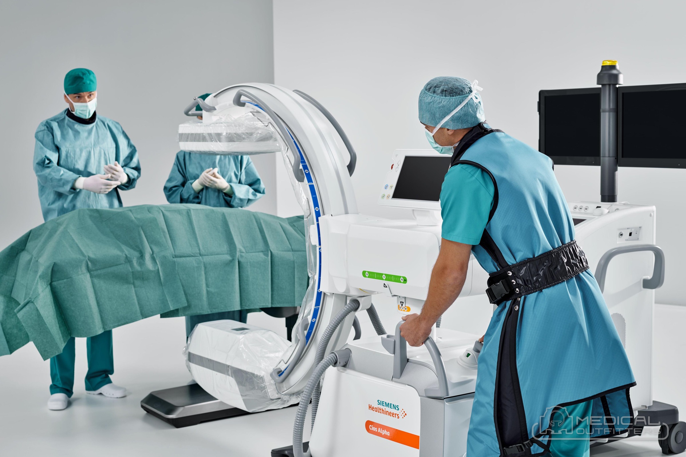
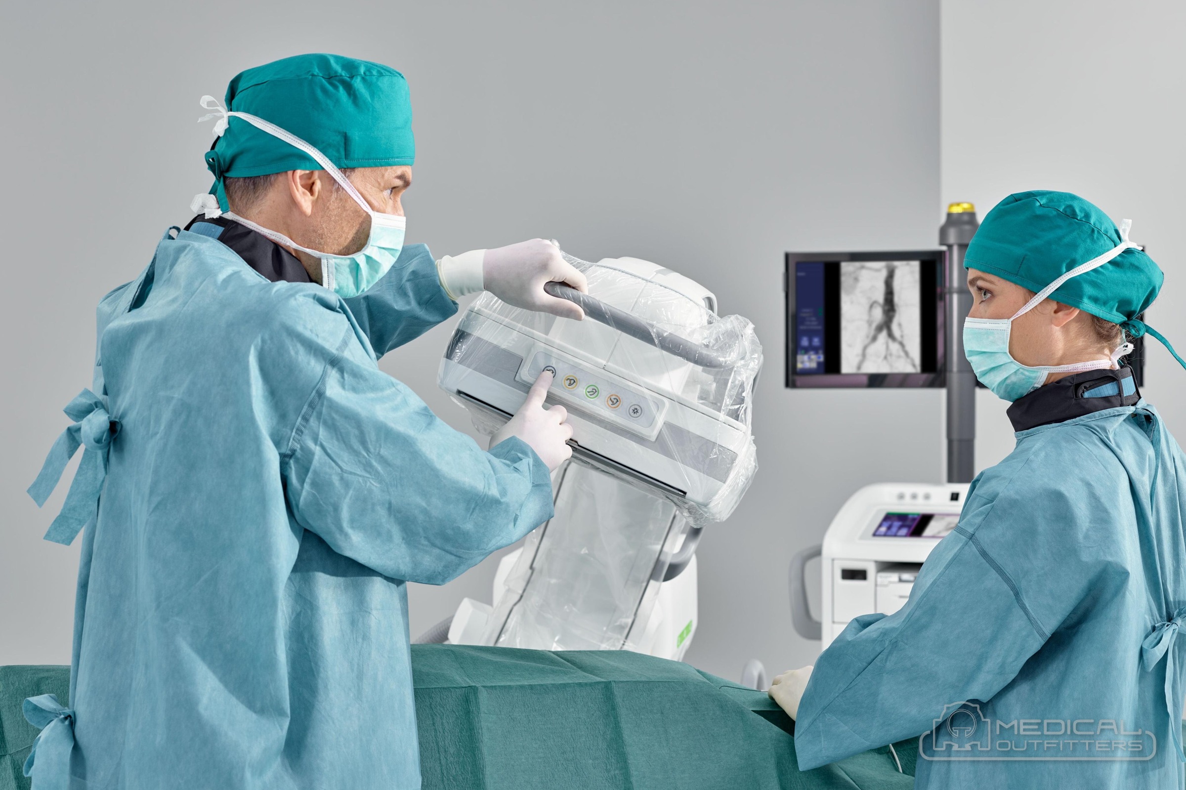
Who runs it varies by state and by setting. In a hospital operating room a radiologic technologist usually drives the C-arm while the surgeon controls the exposures with a foot pedal or asks the technologist to fire; in a pain practice the physician often works with a technologist at the side. State radiation control programs set who may operate fluoroscopy equipment, so a facility adding a C-arm checks its state’s operator and registration rules before the machine arrives.
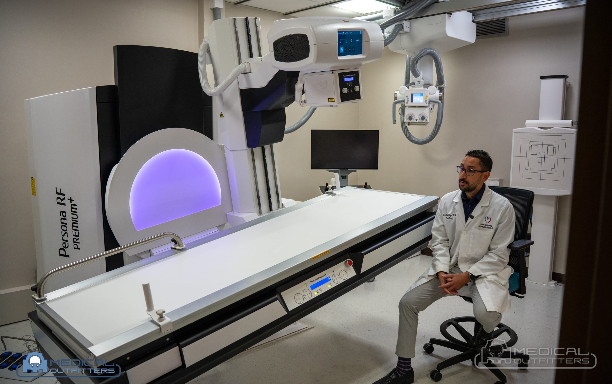
The C-arm’s mobility is what makes it a better fit than a fixed room for this work. It can be wheeled from one operating room to the next, moved out of the way when it is not needed, and set up in minutes. A fixed radiography and fluoroscopy room, by contrast, is built into radiology for barium studies, swallow studies, arthrograms and myelograms, and is a construction project with shielding designed by a physicist. Our C-arm vs fluoroscopy comparison walks through which practice needs which, and our project post on a Fujifilm Persona RF fluoroscopy room in Puerto Rico shows what the fixed alternative involves.
C-Arm Radiation Dose: The Limits, the Alarms and the Habits That Matter
Because fluoroscopy can run for minutes rather than milliseconds, it can deliver more radiation than a plain X-ray. The FDA states that fluoroscopy can result in relatively high doses, especially in complex interventional procedures, and advises using the lowest acceptable exposure for the shortest time necessary. Federal law sets how the machine itself must behave, through the performance standard at 21 CFR 1020.32, which the FDA revised in 2005 to improve the dose information shown to physicians.
What the federal standard requires of the machine:
- Dose rate ceiling: equipment made on or after May 19, 1995 may not exceed an air kerma rate of 88 mGy per minute in normal mode, or 176 mGy per minute in high-level control mode, under 21 CFR 1020.32(d).
- Minimum distance to the skin: the tube must stay at least 30 cm from the patient’s skin on mobile fluoroscopes and 38 cm on stationary ones, with shorter limits only for specific surgical uses and never under 20 cm, under 21 CFR 1020.32(g).
- Dose display: systems made on or after June 10, 2006 must show the air kerma rate and the cumulative air kerma at the operator’s working position, under 21 CFR 1020.32(k).
- The five-minute alarm: the same systems must sound an audible signal for every five minutes of fluoroscopy time, under 21 CFR 1020.32(h).
What the hospital is accountable for. The Joint Commission treats prolonged fluoroscopy with a cumulative peak skin dose above 15 Gy (1,500 rad) to a single field as a sentinel event, as Stanford University’s radiation protection guidance summarizes, and accredited hospitals must review any such case. In practice the cumulative air kerma on the C-arm display is the number a physicist uses to estimate the skin dose, which is one reason a pre-2006 system without that display is harder to defend in an accredited setting.
The habits that cut dose for patient and staff: use pulsed fluoroscopy at the lowest workable pulse rate; collimate the beam to the area of interest; keep the detector as close to the patient as possible and the tube as far away as the arm allows; keep the tube under the table so scatter goes toward the floor; use last-image-hold instead of re-exposing; and step back, because scatter falls off quickly with distance. Staff wear lead or lead-equivalent aprons, thyroid shields and dosimeters, and many rooms add ceiling-mounted shields. A medical physicist surveys the machine when it is installed and as the state requires afterward, which is part of the running cost covered in our cost guide.
What a C-Arm Machine Costs to Buy or Rent in 2026
A C-arm machine costs what its format, detector and condition say it costs. The Medical Outfitters C-arm machine cost guide publishes the ranges below for a system delivered, installed and warrantied, with applications training included, which is why they read higher than equipment-only prices found elsewhere.
| C-arm type | Refurbished | New | Rental per month |
|---|---|---|---|
| Mini or extremity C-arm | $45,000 to $85,000 | $110,000 to $185,000 | $2,000 to $4,500 |
| Full-size mobile C-arm | $55,000 to $135,000 | $165,000 to $295,000 | $2,500 to $7,000 |
| 3D or premium vascular C-arm | $125,000 to $240,000 | $275,000 and up | $5,000 to $12,000 |
Purchase ranges are from the Medical Outfitters C-arm cost guide; rental ranges are from the Medical Outfitters C-arm rental page, delivered, set up and supported for the term. The cost guide states that a certified refurbished C-arm saves 40 to 60 percent against new with comparable image quality, provided the tube, detector and generator were tested to specification.
Where a quote lands inside the band depends on the detector (a flat panel costs more than an intensifier), the generator power, the tube’s history, the arc geometry and motorization, the licensed software, and the warranty. A refurbished GE OEC 9900 Elite, for example, runs $85,000 to $160,000 depending on its configuration, per our cost guide, while a refurbished OEC 9800 sits in the lower half of the full-size band.
Rent or buy. Renting fits a need with an end date: a system out for repair, a second room for a few months, or a new service line with unproven volume. The C-arm rental page works the arithmetic: at full-size rental rates, about nineteen to twenty-two months of rent equals the price of a refurbished system, and buying usually wins past about a year of steady use.
Running costs include the service contract, tube and detector repairs, the physicist’s survey and state registration, lead aprons and shields, and on some current platforms annual software subscriptions. Ask for the service contract in writing with the purchase quote. Medical Outfitters services and repairs C-arms, including systems bought elsewhere, through our service and repair team, and for a wider view of where a C-arm sits against other modalities, see radiology equipment cost in 2026.
Choosing and Pricing a C-Arm With Medical Outfitters
A C-arm is the most flexible imaging system a surgical facility can own: one machine on wheels that brings live X-ray to every room that needs it. Choose the format from the case list first (mini for extremities, full-size for the operating room, 3D for spine and trauma checks), then the detector for the years you plan to keep it, then new or refurbished for the budget. Start with the C-arm machine cost guide for the bands by model, read the used C-arm buying guide before comparing used systems, browse C-arm machines for sale, and check the C-arm rental terms if the need has an end date. When you know your procedures and your rooms, send them to us and we will price a specific system.
- US Food and Drug Administration, Fluoroscopy: definition, uses, risks and the performance standard reference: https://www.fda.gov/radiation-emitting-products/medical-x-ray-imaging/fluoroscopy
- 21 CFR 1020.32, Fluoroscopic equipment (Cornell Legal Information Institute text): air kerma rate limits, source-skin distance, extremity C-arm labeling, last-image-hold, dose display and the five-minute signal: https://www.law.cornell.edu/cfr/text/21/1020.32
- Stanford University Environmental Health and Safety, Sentinel event, fluoroscopy or radiotherapy: the Joint Commission 15 Gy peak skin dose threshold: https://ehs.stanford.edu/manual/radiation-protection-guidance-hospital-staff/sentinel-event-fluoroscopy-or-radiotherapy
- Siemens Healthineers, Cios Spin mobile C-arm product page: 30 x 30 cm CMOS detector, 25 kW generator, 400 projections, 30 second scan, 25 x 25 x 16 cm volume: https://www.siemens-healthineers.com/en-us/surgical-c-arms-and-navigation/mobile-c-arms/cios-spin
- GE HealthCare, OEC Elite CFD product page: 21 or 31 cm CMOS detector, arc options and the 22 percent field-of-view comparison: https://www.gehealthcare.com/products/surgical-imaging/oec-elite-cfd
- Medical Outfitters, C-Arm Machine Cost and Buying Guide (2026): purchase bands, models, detectors, generators and running costs: https://medicaloutfitters.com/c-arm-machine-cost-guide/
- Medical Outfitters, C-Arm Rental: Cost, Terms and How It Works (2026): monthly rental bands and the rent versus buy arithmetic: https://medicaloutfitters.com/c-arm-rental/
- C-arm machine cost and buying guide (2026)
- C-arm machines for sale
- C-arm rental: cost, terms and how it works
- Used C-arm buying guide
- C-arm vs fluoroscopy: what is the difference
- Ziehm Vision RFD 3D mobile C-arm
- Ziehm Solo FD compact C-arm
- Orthoscan TAU 1515 mini C-arm
- Fujifilm Persona C mobile fluoroscopy system
- What is a cath lab
- Radiology equipment cost in 2026, every modality
- Imaging equipment service and repairs
- Price a C-arm for your facility
C-Arms: Common Questions
What is a C-arm?
A C-arm is a mobile X-ray machine with the X-ray tube and the image detector at opposite ends of a C-shaped arm. The patient lies between them, and the system shows live X-ray video, called fluoroscopy, so a surgeon can watch bone, implants, needles or contrast during a procedure. The arm rotates and tilts around the table, giving any viewing angle without moving the patient. Mobile C-arms wheel between operating rooms and need no dedicated shielded room.
What does C-arm stand for?
Nothing beyond its shape. The name describes the curved arm, shaped like the letter C, that connects the X-ray tube at one end to the detector at the other. It is not an acronym. People also write it as C arm, C-arm X-ray or C-arm machine, and all of those terms describe the same mobile fluoroscopy equipment used in operating rooms, surgery centers and pain management practices.
How does a C-arm X-ray work?
The generator fires short pulses of X-rays from the tube through the patient. Bone, metal and contrast absorb more of the beam than soft tissue, and the detector at the top of the arm turns what passes through into a video frame. The system repeats this several times a second, adjusts the technique automatically for thicker or thinner anatomy, and shows the result live on a monitor. The last frame stays on screen when the pedal is released.
What is the difference between a C-arm and fluoroscopy?
Fluoroscopy is the method: live X-ray imaging viewed in real time. A C-arm is one of the machines that performs it, a mobile unit with the tube and detector on a C-shaped arm. Fixed radiography and fluoroscopy rooms and cath labs use the same method from a permanent installation in a shielded room. Every C-arm does fluoroscopy, but not every fluoroscopy system is a mobile C-arm.
What is a mini C-arm?
A mini C-arm is a small, low-power C-arm for the extremities: hands, wrists, elbows, feet and ankles. It has a short arm and a small flat detector, such as the 15 x 12 cm to 20 x 20 cm panels on the Orthoscan TAU family, and the lowest dose in the category. Under 21 CFR 1020.32, short C-arms made since June 10, 2006 must be labeled for extremity use only, so a mini cannot image a spine or hip. Every mini we sell new or supply pre-owned is priced in our mini C-arm price guide.
What is a C-arm used for?
C-arms guide orthopedic and trauma surgery such as fracture fixation and joint replacement, spine surgery and screw placement, pain management injections and nerve ablations, urology procedures for kidney stones and stents, and, on higher-power systems, vascular, cardiac and GI work such as angiography, lead placement and ERCP. The FDA lists catheter guidance, stent placement, angiography and orthopedic surgery among the main uses of fluoroscopy.
Is C-arm radiation dangerous?
The FDA calls the chance of harm from a given procedure statistically very small, but long cases can deliver high doses, so the machine and the habits both matter. Federal rules cap the dose rate at 88 mGy per minute in normal mode and require a dose display and a five-minute alarm on systems made since 2006. Pulsed fluoroscopy, tight collimation, the detector close to the patient and lead aprons for staff keep exposure low.
How much does a C-arm cost?
Per the Medical Outfitters C-arm cost guide, a mini C-arm costs $45,000 to $85,000 refurbished and $110,000 to $185,000 new, a full-size mobile C-arm $55,000 to $135,000 refurbished and $165,000 to $295,000 new, and a 3D or premium vascular system $125,000 to $240,000 refurbished or $275,000 and up new, delivered, installed and warrantied. Renting costs $2,000 to $12,000 a month by class.
Related Articles