Request a QUOTE!
Better systems, better service, and better healthcare are at your reach.
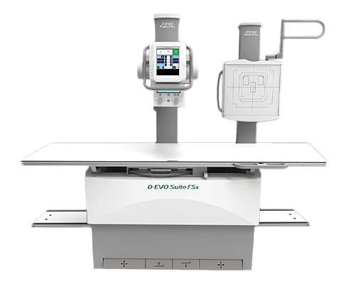
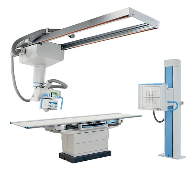
Overview
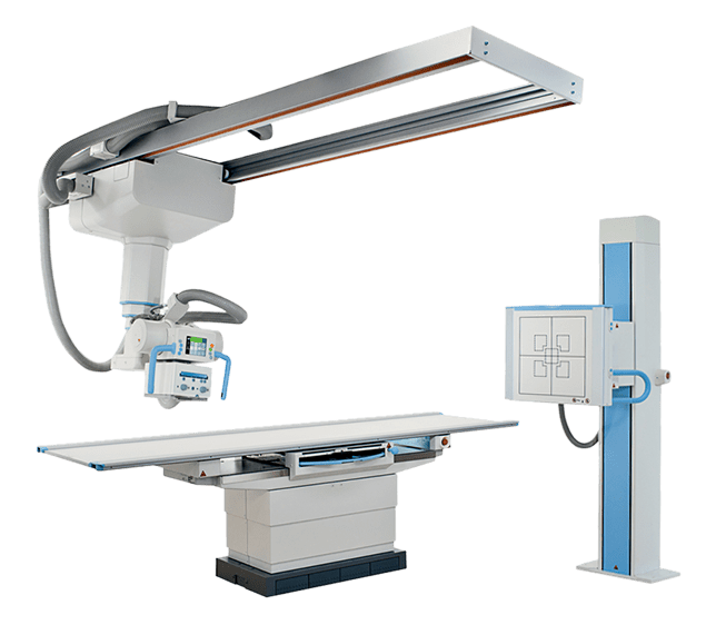
Take full advantage of digital radiography workflow in an x-ray room that is optimized for use with FDR D-EVO detectors. D-EVO Suite II offers just the right balance of automated and manual movements for the grab-and-go speed and dependability needed for even the most demanding environments.
Helping the technologist take control
D-EVO Suite II is designed to put the technologist in full control of the system while staying close to the patient on the table or at the chest stand. The lightweight, ceiling mounted x-ray tube allows lighter and smoother positioning. The tube head features new automation and display features, including a convenient touchscreen for system adjustments traditionally found at the generator console. The system’s workflow and auto tube tracking enhancements also provide benefits in areas where speed during the exam is critical.
Any of Fujifilm’s FDR D-EVO II, III or FDR ES detectors, including the 17×17” detector can be used interchangeably with the D-EVO Suite II. New D-EVO III detectors are even more lightweight and more rugged. Multiple sizes allow technologists to mix, match and share sizes and capture types throughout the radiology department, including other rooms and portables.
These units are to be sold installed as turn-key projects.
ISS and Dynamic Visualization™
The D-EVO Suite II uses two of Fujifilm’s innovations: Irradiated Side Sampling (ISS) and Dynamic Visualization.
Fujifilm’s patented Irradiated Side Sampling (ISS) technology increases sharpness and DQE by reducing light scatter and blur. Fujifilm positions its thin-film transistors (TFT) at the top of the capture layer, reading image data where it is sharpest and strongest, resulting in exceptional detail and image quality, even at lower doses.
Fujifilm’s Dynamic Visualization image processing automatically recognizes the region of interest and applies the optimum image processing parameters throughout the entire exposure field, dynamically extending contrast visibility, and improving window and leveling for exceptional images with high diagnostic detail. Additional advanced functions include:
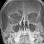
- Multi-Frequency Processing (MFP): Applies edge enhancement and gray scale processing to multiple frequencies, improving visibility for varying densities and foreign structures. Useful in viewing spine, skull and orthopedic hardware images
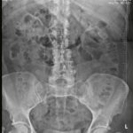
- Flexible Noise Control (FNC): Selectively suppresses noise without loss of sharpness. Useful for pediatrics, lumbar spine and abdomen views
- Grid Pattern Removal (GPR): Intelligently removes moiré patterns caused by grids
- Fujifilm DR to Fujifilm Synapse PACS viewing shortcuts: Applies radiologist-specific single-click processing preferences to views, significantly simplifying workflow
- Auto-stitching is optionally available for scoliosis and long-leg images
Auto Stitching for long length imaging
Automated long length image capture at the chest stand is available as an option. Synchronized detector and tube travel rapidly acquires up to 3 images which are intelligently stitched together by the workstation software for one seamless long length image. Option needs to be purchased with the room up front and includes detector built into the chest stand, motorized tube angulation movements and image stitching software.
- Multi-Frequency Processing (MFP): Applies edge enhancement and gray scale processing to multiple frequencies, improving visibility for varying densities and foreign structures. Useful in viewing spine, skull and orthopedic hardware images
- Alignment markers on the patient positioning shield/stand assist software to accurately align images
- Seamlessly stitches multiple images together using FDX Console software, which automatically recognizes specially-fixed markers
- Motion Suppression intelligently adapts image alignment to reduce effects of patient movement between exposures


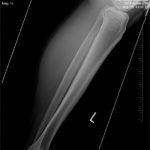


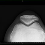


ISS Technology
Axillary View Off-centering
KUB Bariatric
Sunrise View
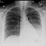
AP Chest Large Male
Swimmer’s View Trauma
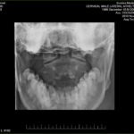
Lateral Cervical Spine
Pediatric Elbow Off-centering
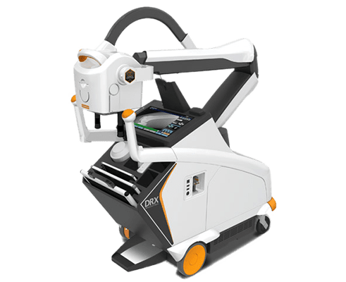
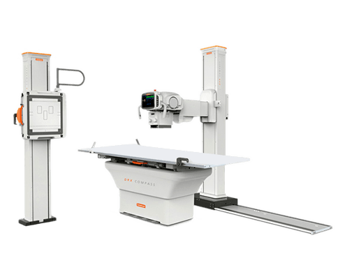
Related Products