Request a QUOTE!
Better systems, better service, and better healthcare are at your reach.
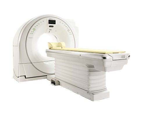
Overview
In radiation oncology, successful outcomes are measured by consistently and confidently moving patients into the right treatment plan. Which is where CT technology can help—by delivering accurate and reliable data for treatment planning for every cancer patient.
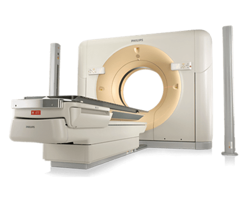
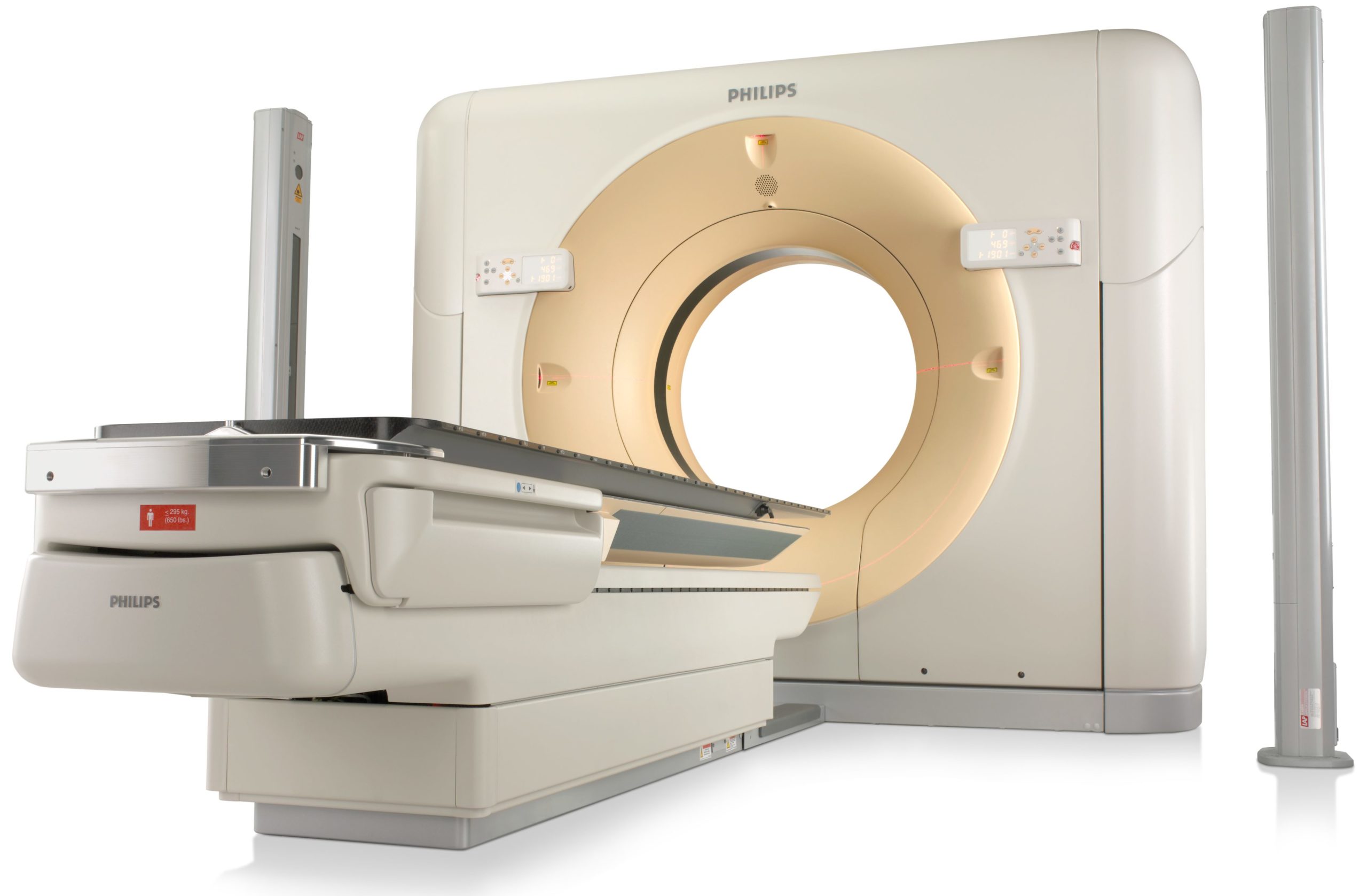
As the first CT scanner specifically designed for radiation oncology, Philips CT Big Bore delivers accuracy, superb imaging performance, an intuitive workflow, and quantitative integrity— empowering you to deliver precise treatment plans for your patients.
These units are to be sold installed as turn-key projects.
- Accuracy: 60cm scan field of view (SFOV) Ensures that image quality and Hounsfield Unit accuracy is maintained out to 60cm— critical to calculate tissue density in planning.
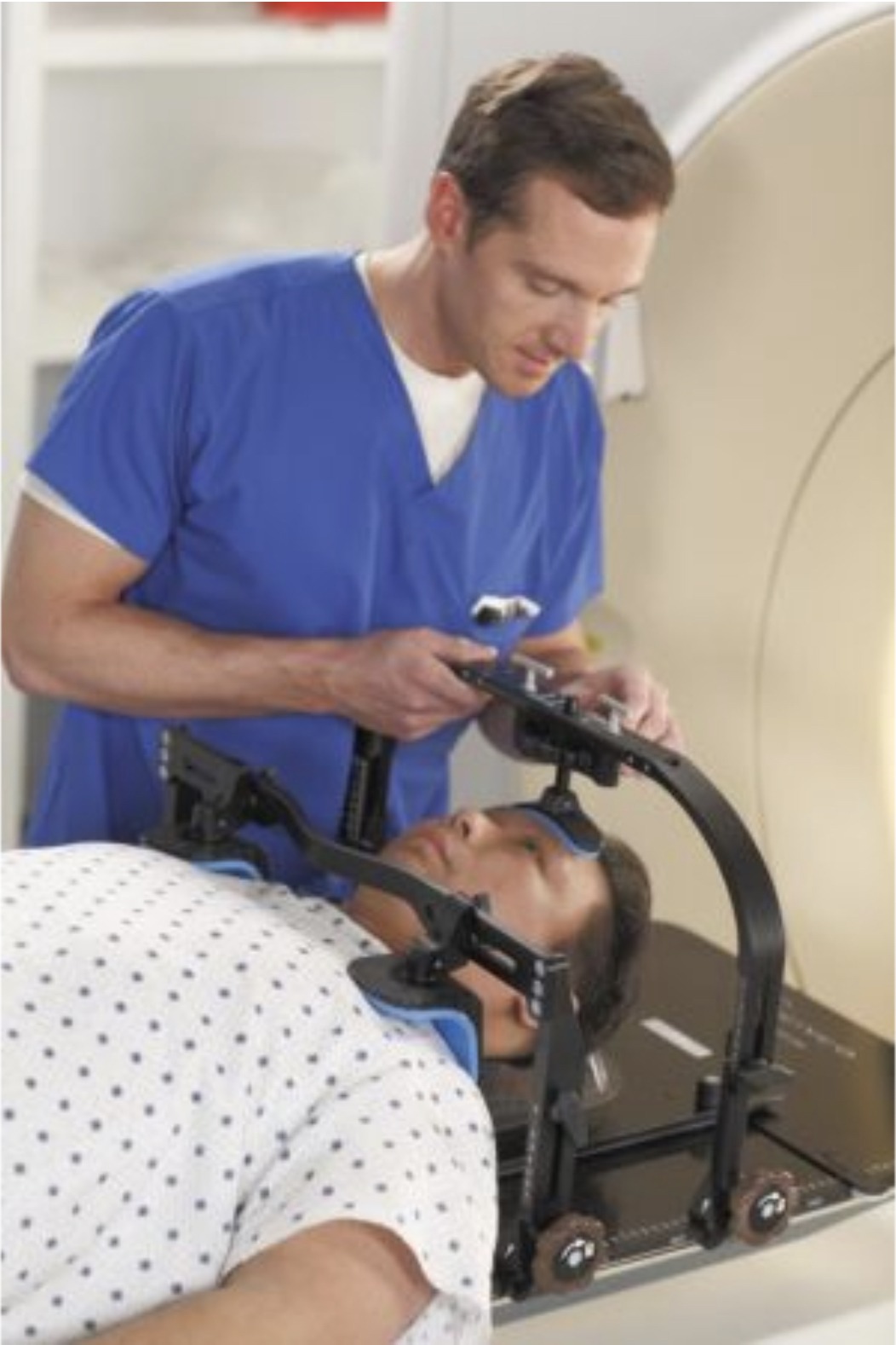
- Patient positioning: 85cm patient aperture Accommodates even complex patient setups and allows for imaging the patient in the optimal treatment position for accurate treatment planning.
- Imaging performance: iDose4 and O-MAR iDose4 improves image quality* through artifact prevention, noise reduction, and increased spatial resolution at low dose. O-MAR reduces artifacts caused by large orthopedic implants and improves visualization of organs and structures. Together, they produce high image quality with reduced artifacts.
- Motion evaluation: 4D CT tools Evaluate motion of the tumor and critical organs during breathing to aid in making clinical decisions regarding the size of the target volume and gated treatment delivery. The resulting images are used to identify a target volume that encompasses the entire range of tumor motion.

iPatient
Spend more time with the patient and less overall time on the procedure. Delivering dedicated oncology ExamCards, this advanced platform provides a standardized, patient-centered approach to imaging simulation with consistency from scan-to-scan.
Pulmonary Toolkit for Oncology
To evaluate how a tumor moves through the patient’s breathing cycle, this comprehensive set of tools helps improve accuracy in treatment planning and therapy delivery, even in patients who have difficulty breathing regularly.
TumorLoc
This application provides fast and efficient tools for patient marking and simulation directly from the scanner console. This includes consistent and reproducible setup from simulation to treatment, the ability to localize isocenter for treatment targets, and palliative “Sim to Treat” tools that bypass simulation and go directly to treatment. Accurate and efficient workflow powered by the Pinnacle treatment planning system
Configuration
- Therapy planning table top
- 8.0 MHU MRC X-ray tube
- 16 x 0.75mm isotropic imaging
- True 3-D cone beam reconstruction
Dual monitor configuration - Oncology specific protocols
X-ray system
- MRC X-ray tube with maximum heat anode storage of 8.0 MHU
- Dynamic focal spot
- 60kW generator system Data acquisition system
- 16-slice detector configuration
- 24 mm volumetric coverage Patient support
- 650 lb. (295 kg.) table capacity with full Z accuracy
Dose management
- DoseRight ACS (Automatic Current Selection)
- DoseRight DOM (Dynamic Dose Modulation)
- DoseRight ECG-gated dose modulation


Scan specifications
- True 60 cm scan field of view
- 16 x 0.75 isotropic imaging
- Sub second rotation time: 0.44, 0.5, 0.75, 1.0, 1.5, 2.0 secs
- Sub-millimeter slice width 0.60mm
- RapidView reconstruction
- Recon matrix up to 5122, 7682 and 10242
- True 3-D cone beam reconstruction
Image specifications
- Spatial resolution: 15 Lp/cm maximum at 0% MTF (axial and spiral)
- Low contrast 4mm at 0.3% measured on 20cm CATPHAN phantom
Related Products