
Request a QUOTE!
Better systems, better service, and better healthcare are at your reach.
Overview
The Hologic Discovery DXA Bone Densitometry system enables you to identify patients at risk for osteoporosis and other debilitating conditions. Delivering exceptional precision and pinpoint accuracy, Hologic Discovery DXA technology delivers high-quality images for your at-risk patients.
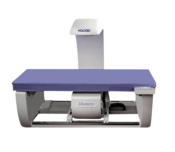
The Discovery DXA system features bone densitometry technology that allows clinicians to assess bone health in patients who may be at risk.
Discovery delivers:
- High-resolution digital detector array, which improves fracture detection visualization of abdominal aortic calcifications.1
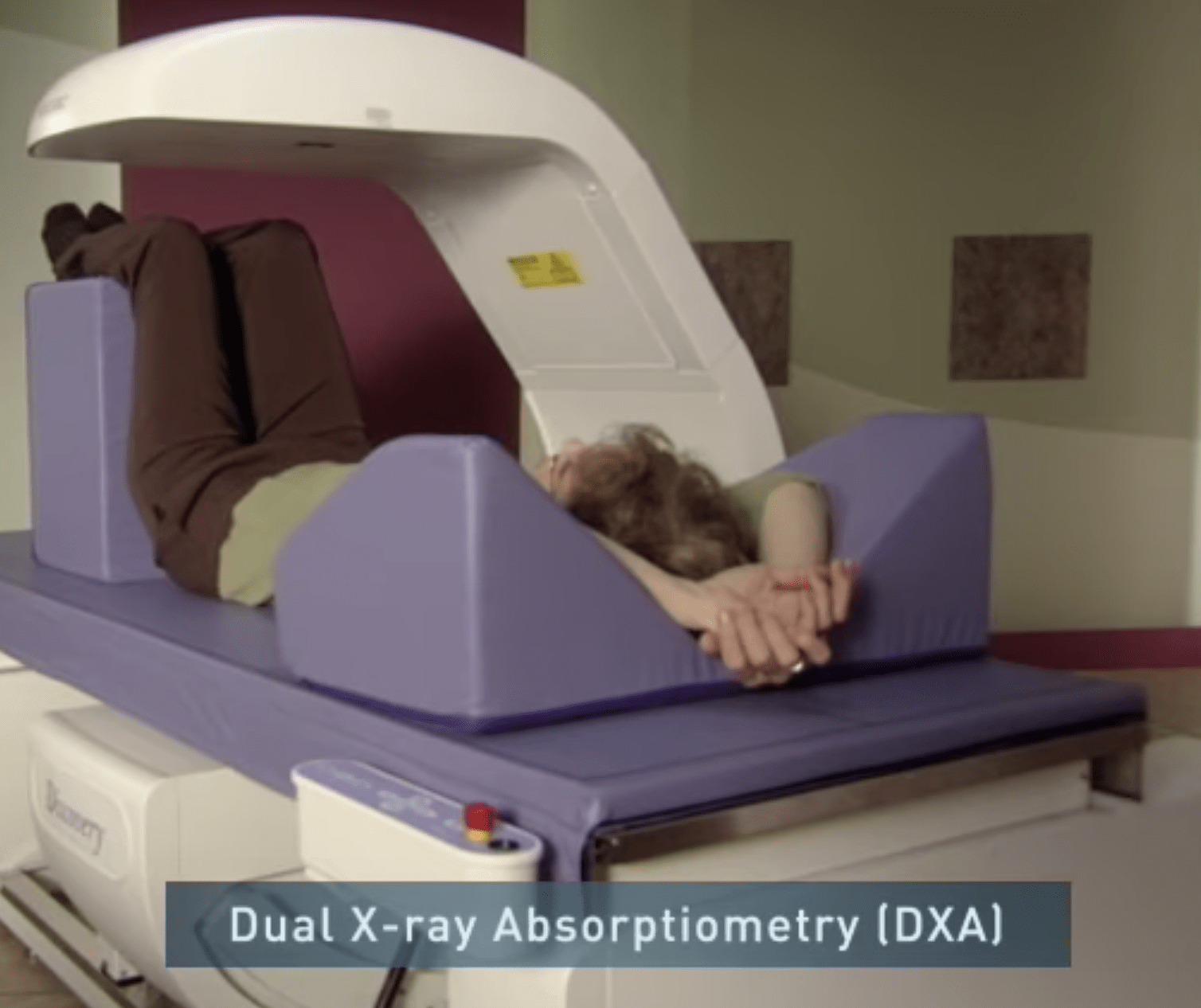
- Speed and image quality, with images of the hip and spine captured in as fast as 10 seconds.
- Superb visualization, an exclusive Hologic design that utilizes a high-resolution detector array paired with true fan-beam linear acquisition geometry.1
- Continuous automatic calibration, ensuring precise and consistent measurements results from exam to exam.
The Discovery DXA system offers exceptional precision and accuracy to help you identify patients with osteoporosis and/or who are at risk for fractures.

Features of the Discovery DXA system include:
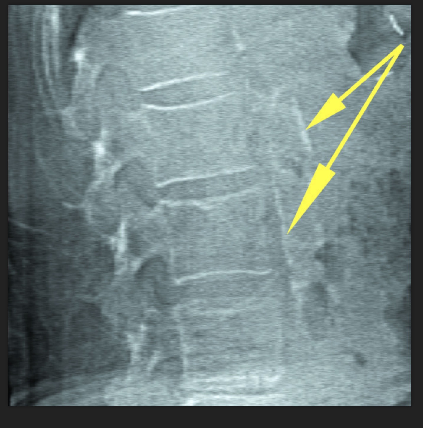
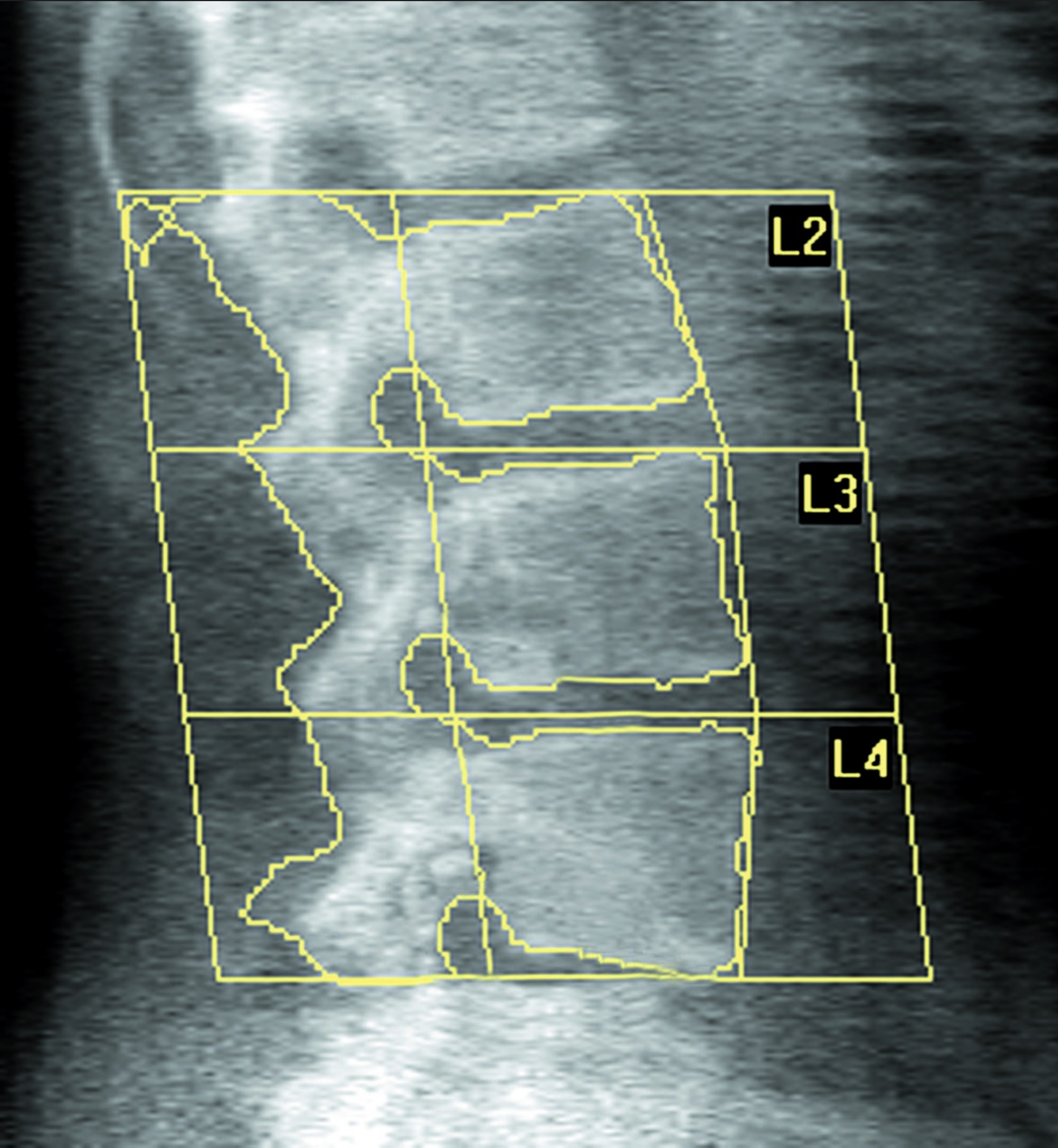
High Definition Instant Vertebral Fracture Assessment (IVA-HD)
The IVA-HD tool dramatically improves the detection of vertebral fractures by doubling the resolution of previously available techniques with a low-dose, single-energy image. Vertebral fracture status, combined with traditional bone density testing, is the gold standard for fracture risk assessment.2
Abdominal aortic calcification
The IVA-HD tool also allows you to visualize calcified plaques in the abdominal aorta, which may be a significant indicator of heart disease and stroke.3-5 Exclusive to Hologic, this feature provides vital information that may link to coronary heart disease (CHD), which is the leading cause of death for women. In fact, nearly 39 percent of all female deaths in the U.S. are due to CHD.6
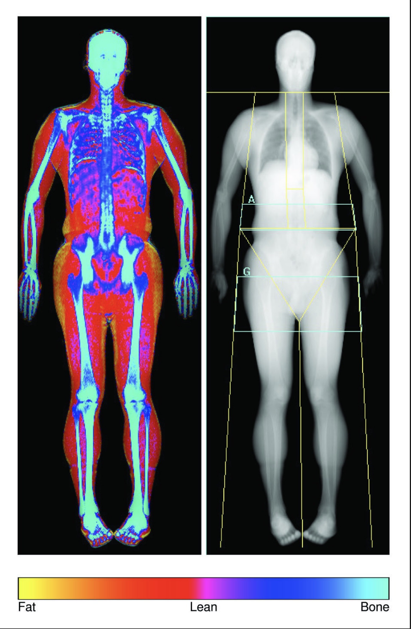
Advanced Body Composition Assessment and Visceral Fat Assessment
The Advanced Body Composition assessment feature produces color images displaying the distribution of fat, lean mass, bone and fat mass index. The information is translated into an easy-to-interpret report for improved patient management and counseling.
Our DXA systems incorporate the National Health and Nutrition Examination Survey (NHANES) whole body composition reference data. The Endocrine Society recommends annual bone mineral density and body composition exams for bariatric surgery patients.7
FRAX* 10-year Fracture Risk Assessment
The FRAX fracture risk assessment tool calculates a patient’s 10-year probability of hip and other osteoporotic fractures. FRAX was developed by the World Health Organization (WHO) to help healthcare providers identify and proactively treat patients with a high risk of debilitating bone fractures due to low bone mass and other significant risk factors.
*FRAX is a trademark of the World Health Organization Collaborating Centre for Metabolic Bone Diseases, located at the University Of Sheffield Medical School, UK.
Reporting Solutions
Whether you are entering patient data or interpreting patient results, Hologic offers comprehensive tools to streamline bone densitometry workflow to improve your practice’s efficiency, giving you more time to spend with patients.
Physicians Report Writer DX is a comprehensive tool that combines the patient’s information, BMD, IVA-HD and FRAX data into an automated electronic report.


Our Discovery DXA system offers some of the latest innovations in bone densitometry technology, including:
- OnePass single-sweep scanning, which delivers superb image quality8 and precision.9 Our exclusive design utilizes a multi-element digital detector array paired with true fan-beam acquisition geometry, enabling rapid, dual-energy bone density measurements. OnePass scanning is designed to eliminate beam overlap errors and image distortion found in rectilinear acquisition techniques, resulting in superb image quality and data stability.

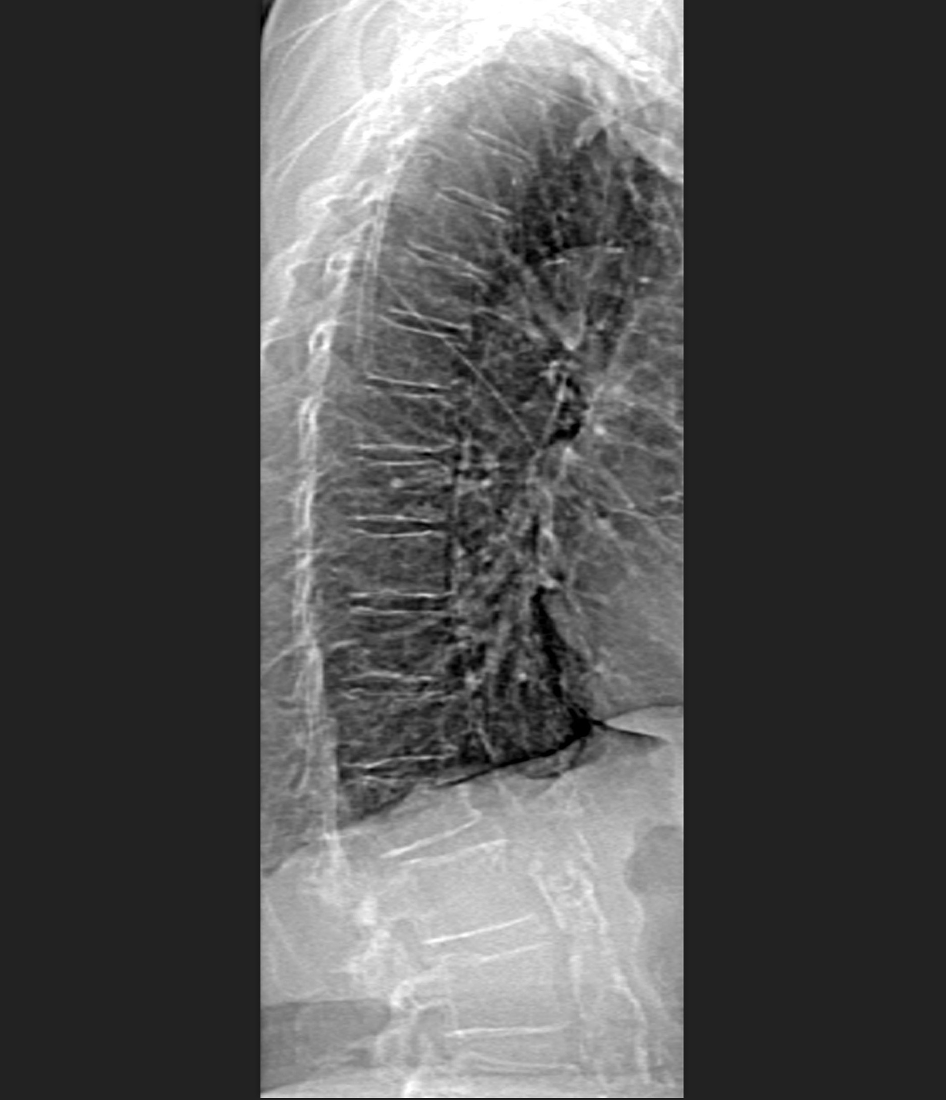
Abdominal Aortic Calcifications
Color Tissue Mapping
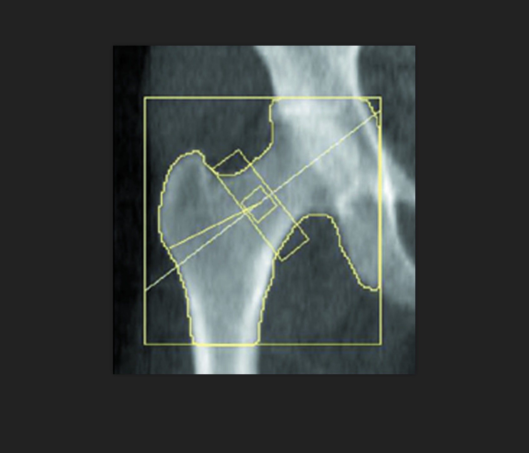
Hip
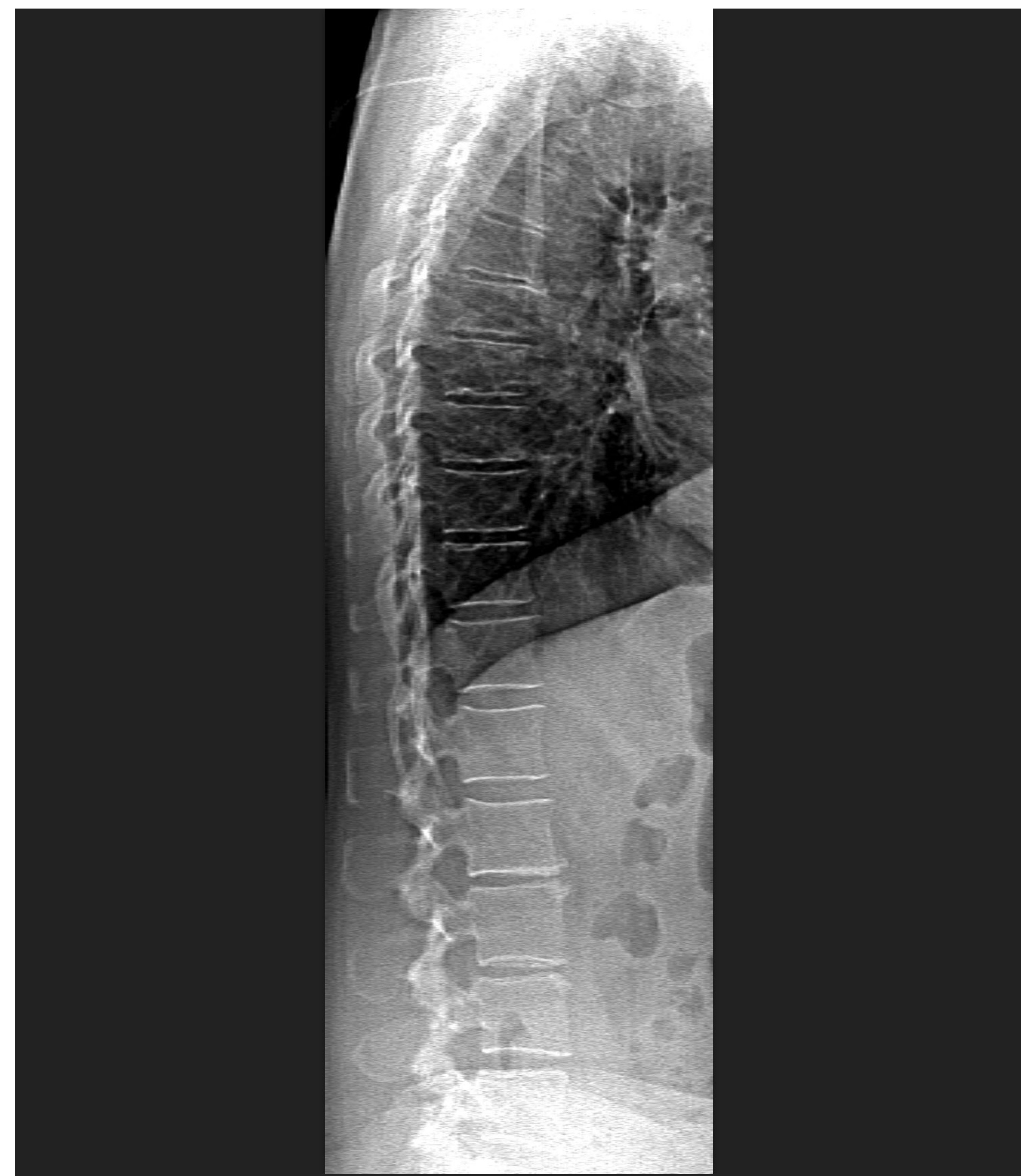
IVA-HD with Fracture
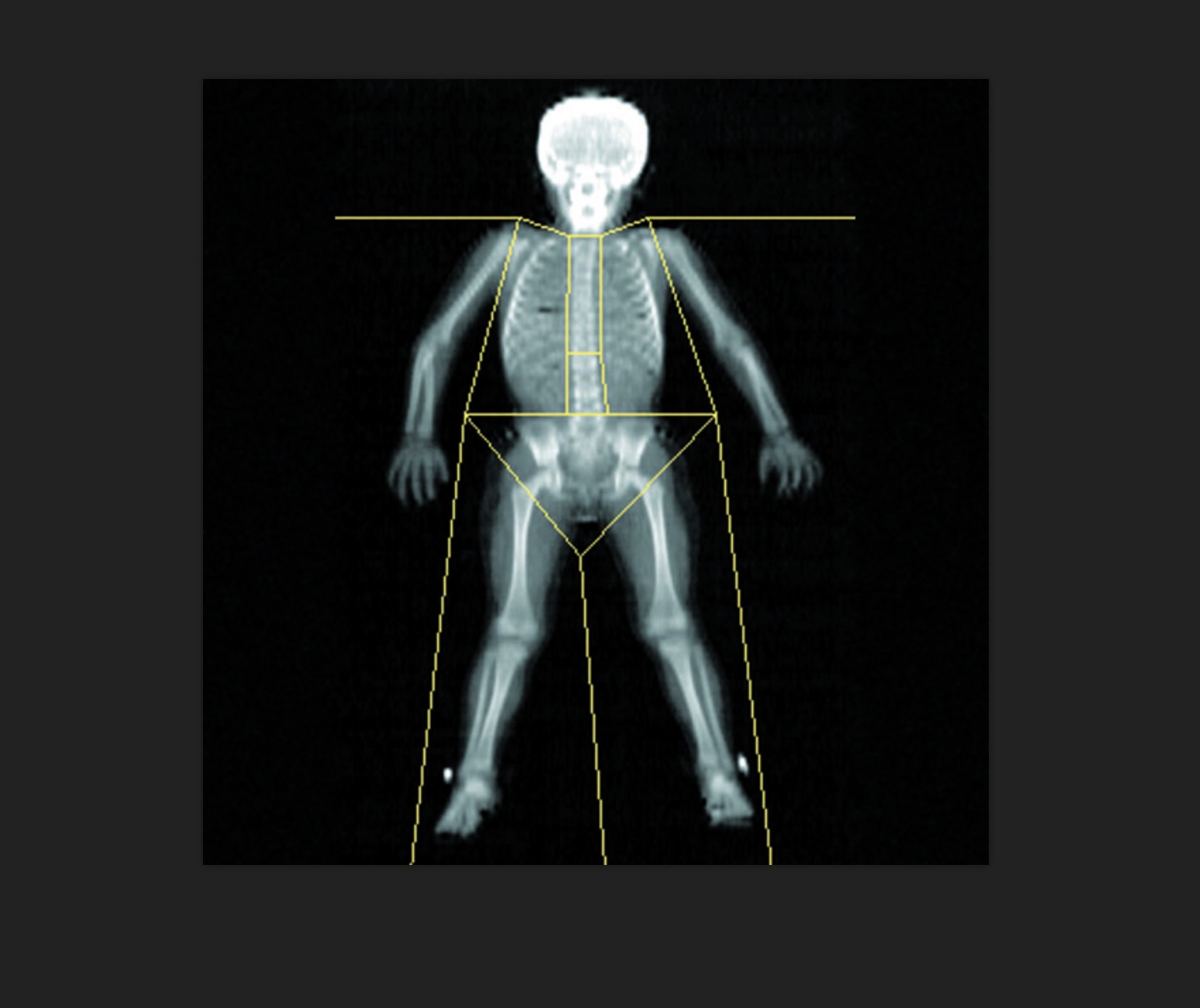
Pediatric Whole Body
IVA-HD
Prosthetic Hip

Small Animal Option

Supine Lateral BMD
Related Products


