What is a PET scan machine, and why does nearly every new one arrive with a CT scanner bolted to the front of it? A PET scan machine is the scanner that turns a tiny amount of injected radioactive tracer into a three-dimensional map of metabolism: where cells are burning sugar fastest, where a prostate cancer marker has collected, where heart muscle is still alive. It is the most important tool in cancer staging and one of the most demanding machines a facility can install, because the scanner is only the middle of a system that also includes a tracer supply with a two-hour clock, shielded rooms and a radioactive materials license. The terms get mixed up constantly: PET scanner, PET scanner machine, PET/CT scanner and PET CT scan are used for the same equipment. This guide explains what the machine is, how a PET scanner works from tracer to image, the parts inside the gantry, what the CT half adds, the tracers it runs on, what the room needs and what a PET scan machine costs in 2026.
Medical Outfitters sources, compares and installs refurbished PET/CT systems from GE HealthCare, Siemens Healthineers, Philips, Canon and United Imaging, and supplies the Mammotome Neoprobe gamma detection system, according to our PET/CT scanner cost guide. We wrote this for the hospital administrator, imaging center owner, radiology manager and clinician who needs to understand the machine before a board approves the project. If you are already comparing systems, our PET/CT scanner buying guide takes the purchase decisions in order.
- What it is: a ring of detectors that records the gamma rays from an injected tracer and reconstructs a 3D map of metabolic activity; today it is almost always paired with a CT in one gantry.
- How it works: each positron from the tracer meets an electron and produces two 511 keV photons that fly apart in opposite directions; the ring counts the pairs that arrive together and traces the line between them.
- The parts: scintillator crystals, light sensors (photomultiplier tubes or silicon photomultipliers), coincidence electronics, the CT half, the patient table and the reconstruction computers.
- The tracer: FDG has a half-life of about 110 minutes, so it is delivered daily from a radiopharmacy with a cyclotron rather than stored on site.
- The cost: $200,000 to $1,500,000 refurbished and $500,000 to $4,000,000 new, delivered, installed and warrantied, per the Medical Outfitters PET/CT scanner cost guide.
What Is a PET Scan Machine? The Scanner Behind the Name
PET stands for positron emission tomography. “Positron emission” describes the signal: the tracer injected into the patient is labeled with a radioactive atom that emits positrons as it decays. “Tomography” describes the output: the machine reconstructs that signal into cross-sectional slices and a full 3D volume, the same way CT reconstructs X-ray data. RadiologyInfo, the patient site of the American College of Radiology and the Radiological Society of North America, describes PET as a nuclear medicine exam that uses small amounts of radioactive tracer to evaluate how organs and tissues are functioning, rather than what they look like.
That is the key difference from every other scanner in a radiology department. CT, MRI, X-ray and ultrasound send energy into the body and record what comes back or passes through. A PET scanner sends nothing into the patient. It is a passive detector: the patient is the radiation source, and the machine’s job is to work out exactly where inside the body each signal started.
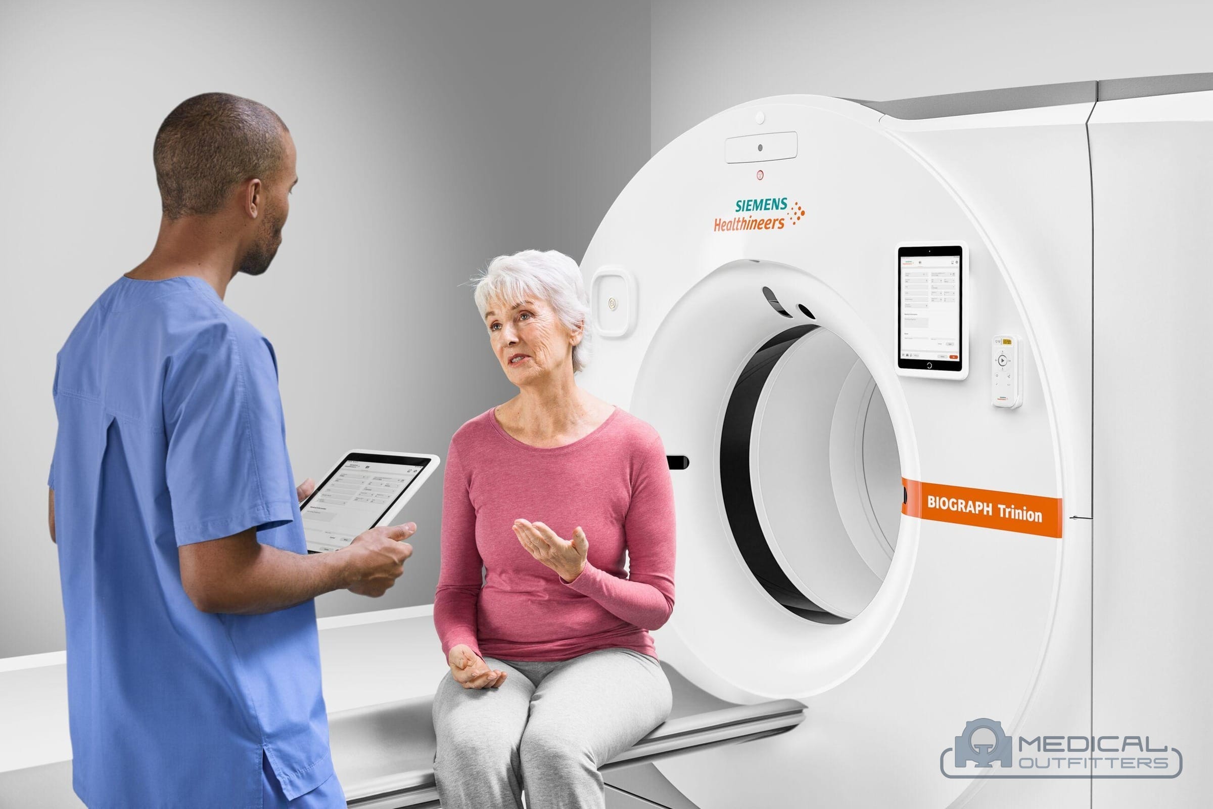
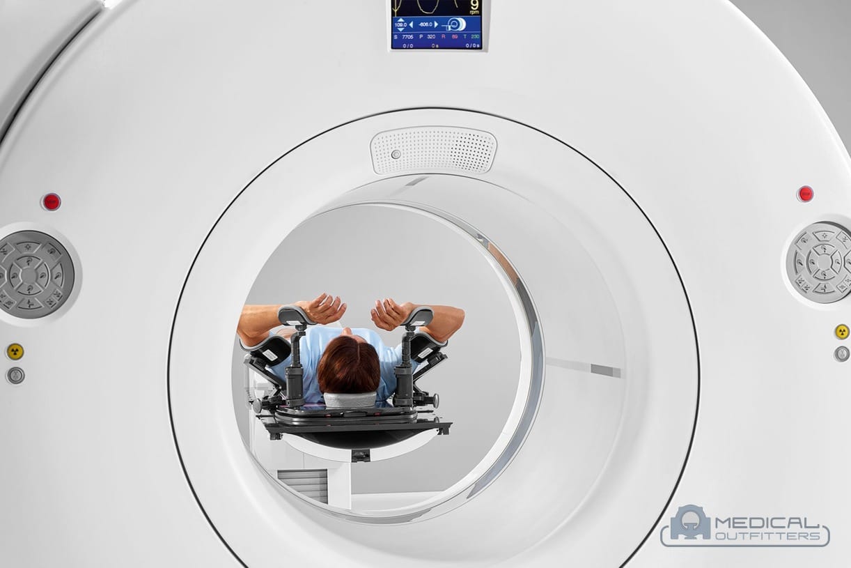
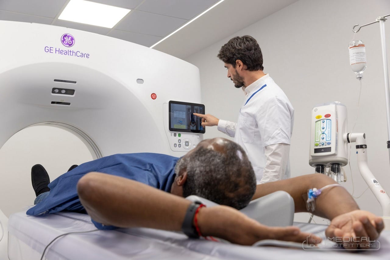
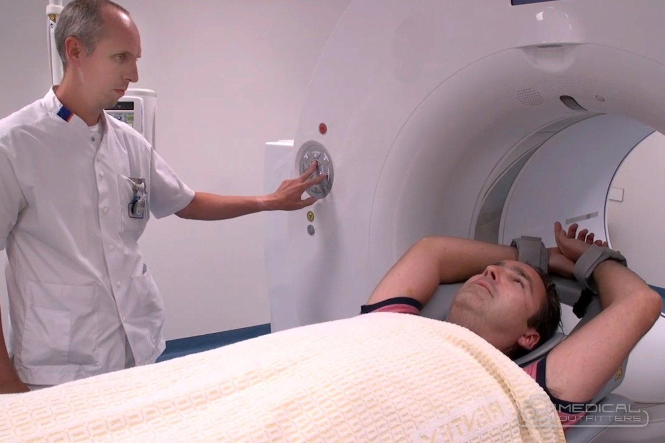
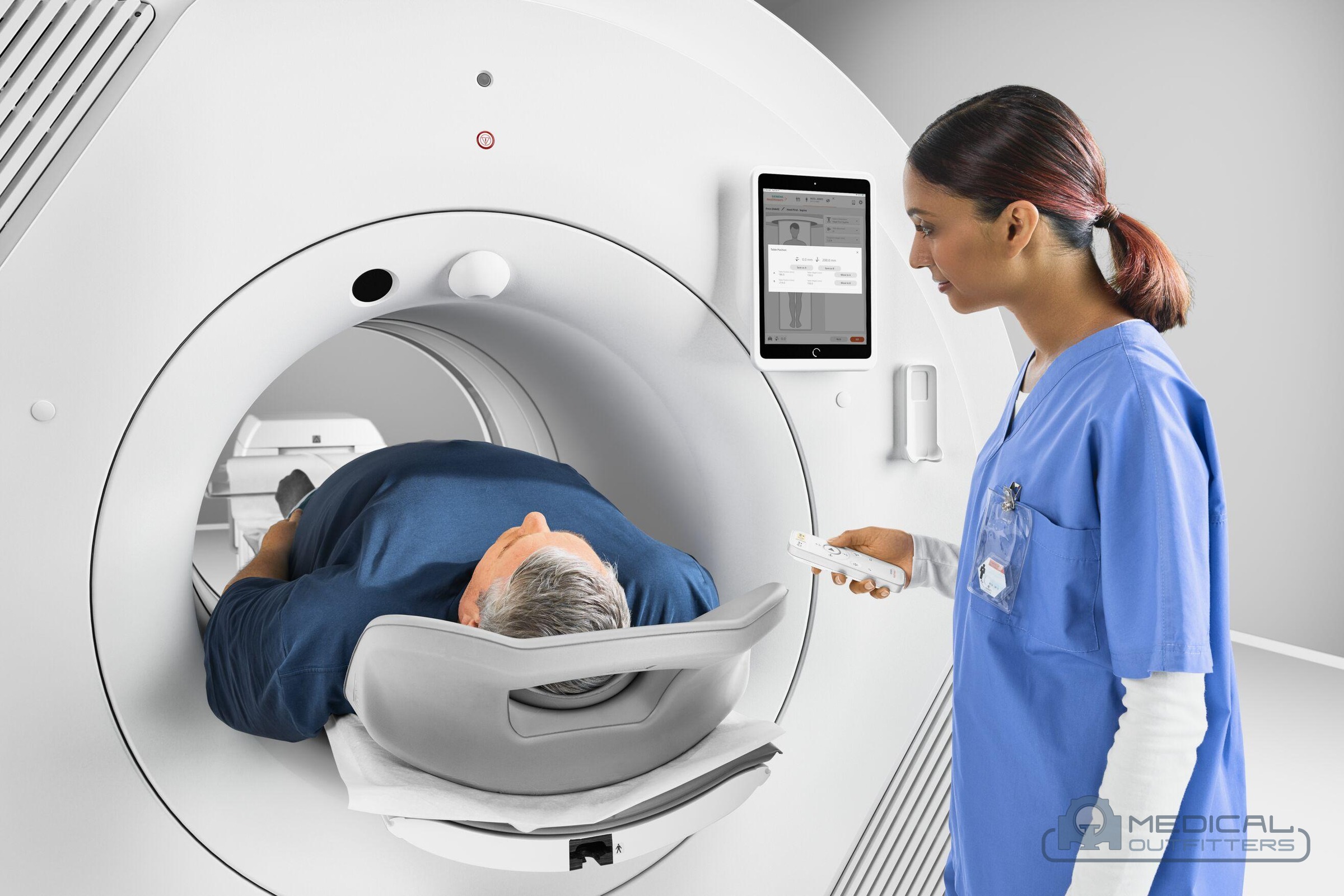
From the outside, a PET scanner looks like a CT scanner with a deeper tunnel. RadiologyInfo describes it as a large, doughnut-shaped machine with multiple rings of detectors inside that record the energy emitted by the tracer. The patient lies on a narrow table that moves through the bore while the rings collect data, and a technologist runs the scan from a control room with a window and an intercom.
PET scanner vs PET/CT scanner. Stand-alone PET scanners still exist in research and in some older installed bases, but they are no longer what hospitals buy. RadiologyInfo states that almost all PET scans today are performed on combined PET/CT scanners. The combination was invented by physicist David Townsend and his colleagues at the University of Pittsburgh: the University of Bristol, which gave Townsend an honorary degree in 2013, records that the first prototype became operational in 1998 and the first commercial PET/CT came to market in 2001, and that TIME magazine named it the medical invention of the year in 2000. A smaller number of PET/MR systems pair PET with an MRI magnet instead. When a buyer says PET scan machine, PET scanner machine or PET CT scanner today, they mean the same thing: a PET/CT.
How Does a PET Scanner Work? From Tracer to Image
A PET image starts with chemistry. The most common tracer, F-18 fluorodeoxyglucose (FDG), is a sugar molecule carrying a radioactive fluorine atom. RadiologyInfo explains that many cancer cells take up glucose faster than normal tissue, so FDG collects in tumors and shows up as bright spots. After the injection, the patient rests quietly while the tracer distributes; RadiologyInfo gives about 30 to 60 minutes for this uptake period, and many protocols run longer.
Annihilation. When an F-18 atom decays, it emits a positron, the antimatter twin of an electron. The positron travels a very short distance in tissue, meets an ordinary electron, and both are destroyed. Their mass turns into two gamma-ray photons of 511 keV each, flying off in almost exactly opposite directions. This is the physics every PET scanner is built around, and the AAPM Task Group 108 report on PET and PET/CT shielding notes that 511 keV photons are far more energetic than the photons used in any other diagnostic imaging.
Coincidence detection. The scanner’s detector ring surrounds the patient. When two detectors on opposite sides of the ring each register a 511 keV photon within a few billionths of a second of each other, the electronics treat the pair as one event and record the straight line between the two detectors, called the line of response. The annihilation happened somewhere along that line. A single photon with no partner is discarded. Over a 20 to 30 minute scan, the ring records hundreds of millions of these lines.
Time of flight. On a time-of-flight scanner, the electronics also measure which of the two photons arrived first. Light travels about 15 cm in a billionth of a second, so a timing difference of a few hundred picoseconds narrows the event to a short segment of the line instead of the whole line. Siemens Healthineers lists a time-of-flight resolution of 214 picoseconds for its Biograph Vision and 178 picoseconds for the Biograph Vision.X. Better timing means less noise in the image, which can be spent on shorter scans, lower injected doses or sharper detail, particularly in larger patients.
Corrections and reconstruction. The raw data are not an image yet. The software corrects for photons absorbed inside the body (attenuation, which is where the CT comes in), for scattered photons, for random coincidences and for the tracer decaying during the scan, then reconstructs slices with iterative algorithms. The final step fuses the PET volume with the CT volume, so the reading physician sees the bright spot of activity laid exactly over the anatomy it sits in.
The Parts of a PET/CT Machine
A PET/CT gantry houses two scanners, one behind the other, with a single patient table that runs through both. The PET half is built from a few components that decide almost everything about image quality and price.
- Scintillator crystals: thousands of small, dense crystals that stop each 511 keV photon and turn its energy into a flash of light. The crystal material sets the detector’s speed and sensitivity. Bismuth germanate (BGO) is dense and sensitive but slow; lutetium oxyorthosilicate (LSO) and its yttrium variant (LYSO) are fast enough for time of flight.
- Light sensors: older scanners read the crystal light with photomultiplier tubes, which are analog vacuum tubes. Digital scanners use silicon photomultipliers (SiPMs), solid-state chips that are faster and can be coupled to small groups of crystals.
- Detector blocks and the ring: crystals and sensors are assembled into blocks, and the blocks into rings. A PET ring is made of a few hundred blocks, 144 to 192 on a Siemens Biograph mCT and 256 on a GE Discovery 690, per the Medical Outfitters PET/CT buying guide.
- Coincidence electronics: the timing circuits that pair photons and, on time-of-flight systems, measure the arrival difference.
- The CT half: an X-ray tube and detector on a rotating ring, usually 16 to 128 slices, described in our guide to what a CT scan machine is.
- Table, consoles and reconstruction computers: a long table that carries the patient through both fields of view, the acquisition console, and the computers that reconstruct and fuse the images.
Axial field of view is the length of the detector rings along the patient, and it decides how much of the body the scanner sees at once. Longer rings catch more photon pairs, so they scan faster or at a lower dose. The three current systems below show how far designs differ.
| Scanner | Detector | Axial field of view | Time of flight | Source |
|---|---|---|---|---|
| Siemens Biograph Vision | 3.2 mm LSO crystals, SiPM | 26.3 cm | 214 ps | Siemens Healthineers product page |
| GE HealthCare Omni Legend 32 | 4.1 mm BGO crystals, SiPM | 32 cm | None (BGO) | Journal of Nuclear Medicine, 2023 |
| Siemens Biograph Vision Quadra | 3.2 mm LSO crystals, SiPM | 106 cm | Yes, Vision detector | Siemens Healthineers, 2021 FDA clearance release |
The Omni Legend shows that the trade-offs are real. A 2023 Journal of Nuclear Medicine evaluation measured its sensitivity at 47.30 cps/kBq at the center of the field, which the authors called class-leading for whole-body PET, while noting that BGO’s timing is unsuitable for time of flight. Siemens announced FDA clearance of the Biograph Vision Quadra on March 8, 2021, with a 106 cm field of view, four times that of the Biograph Vision 600, long enough to image the average patient from head to thigh in one position.
The detector is also the first thing to check on a used scanner. Our cost guide treats detector generation as the single biggest factor in price, ahead of the CT and the brand, and our buying guide covers how block replacement history and daily quality control show a detector’s health.
What Is a PET CT Scan, and Is a PET Scan a CT Scan?
Is a PET scan a CT scan? No. A PET scan detects gamma rays from a tracer inside the body and shows function. A CT scan passes X-rays through the body and shows structure. They are different physics on different detectors. A PET CT scan is both, done back to back on the same table in one visit, without the patient getting up between them, which is why RadiologyInfo says the combined exam can be more accurate than two scans done separately.
The CT does three jobs inside a PET/CT:
- Attenuation correction: the body absorbs some of the 511 keV photons before they reach the ring, more through the pelvis than through the neck. The CT gives a density map of the patient that the software uses to correct the PET counts. Before PET/CT, this took a separate transmission scan with a radioactive rod source that added many minutes.
- Localization: a bright spot on PET alone floats in a blur. Fused with CT, it sits on a named lymph node, a rib or a segment of bowel.
- Diagnostic CT: with 64 slices or more and contrast, the same CT can deliver a full diagnostic study in the same session, or serve as a stand-alone CT when the PET schedule is light.
RadiologyInfo gives the timing: the CT portion takes less than two minutes and the PET portion 20 to 30 minutes, for a total scan time of about 30 minutes, although some exams with additional tracers or drugs extend the visit to as long as three hours.
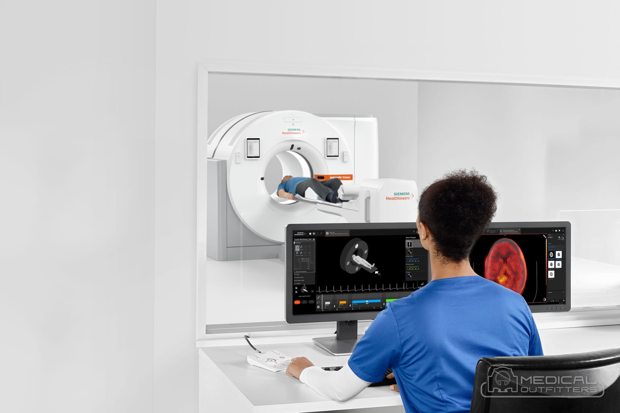
How many CT slices a PET scan machine needs depends on that third job. Sixteen slices are enough for attenuation correction and localization, while 64 or more are worth paying for if the CT will also run diagnostic, cardiac or radiation therapy planning studies, as the Medical Outfitters PET/CT buying guide explains; our guide to CT scanner types by slice count covers what each tier adds.
Dose. Because the exam combines a tracer and a CT, it is one of the higher-dose imaging studies. RadiologyInfo lists about 22.7 mSv for a whole-body PET/CT in an average-sized adult, comparable to 7.6 years of natural background radiation, against about 7.7 mSv for a CT of the abdomen and pelvis and about 3 mSv a year of natural background in the United States. A low-dose CT for attenuation and localization only, and the sensitivity of a newer detector, are the two levers that bring that number down.
PET Tracers: FDG, PSMA and the Two-Hour Clock
A PET scan machine is only as useful as the tracer injected before it. Each tracer is a molecule that goes somewhere specific in the body, labeled with a positron-emitting atom. The half-life of that atom decides how the tracer is made, delivered and scheduled, and every PET department is planned around it.
FDG is the workhorse. Its fluorine-18 label has a half-life of about 110 minutes, according to the AAPM Task Group 108 report and the Medical Outfitters PET/CT cost guide, so a dose loses half its activity roughly every two hours. That is long enough to make FDG in a cyclotron at a regional radiopharmacy and drive it to imaging centers within a few hours, and too short to store. Doses are ordered each scanning day against a confirmed patient list, and a canceled patient is a lost dose.
Prostate-specific membrane antigen (PSMA) tracers have grown fastest. Medicare contractor notices list FDA approval dates for Pylarify (piflufolastat F-18) on May 26, 2021, Illuccix (gallium-68 gozetotide kit) on December 17, 2021, Locametz on March 23, 2022 and Posluma (flotufolastat F-18) on May 25, 2023. The F-18 agents ship like FDG; the gallium-68 kits are labeled on site from a germanium-68/gallium-68 generator, because gallium-68 has a half-life of about 68 minutes.
Cardiac and research tracers push the clock harder. Rubidium-82, used for myocardial perfusion, has a half-life of about 75 seconds, so it comes from a strontium-82/rubidium-82 generator in the scan room, infused directly into the patient on the table. Nitrogen-13 ammonia (about 10 minutes), carbon-11 (about 20 minutes) and oxygen-15 (about 2 minutes) need a cyclotron on site or next door, which is why they are mostly found in academic centers. Amyloid and tau tracers for dementia and fluciclovine for prostate cancer are other F-18 agents delivered the FDG way.
Do you need a cyclotron? No, not for a clinical PET program. The Medical Outfitters cost guide notes that most centers buy tracers per dose from a commercial radiopharmacy with its own cyclotron, and that an on-site cyclotron is a large-network or research decision justified by volume, not by a single scanner. What a site does need is the delivery radius: the buying guide describes FDG supply as a radius on a map around the nearest radiopharmacy, with remote and island sites planning larger doses or air freight.
What a PET Scanner Needs Around It: Shielding, Hot Lab and Uptake Rooms
A CT scanner needs a shielded room. A PET/CT scanner needs a department, because the patient becomes a radiation source from the moment of injection until they leave the building. The AAPM Task Group 108 report treats the injected patient as the source throughout the visit and notes that floors and ceilings, not only walls, may need shielding against 511 keV photons. A CT room rarely converts to PET without added lead or concrete, according to the Medical Outfitters cost guide.
- Scan room: shielding designed by a medical physicist for both the CT X-rays and the 511 keV photons, dedicated three-phase power for the CT, cooling for the gantry and a floor rated for a gantry heavier and longer than a CT’s.
- Hot lab: where doses are received, measured in a dose calibrator and stored behind shielding until injection, with secure access and storage for waste to decay.
- Uptake rooms: quiet, shielded rooms where injected patients rest, typically 45 to 90 minutes per the Medical Outfitters cost guide. Because every patient occupies an uptake room longer than the scan itself, the number of uptake rooms, not the scanner’s speed, usually sets how many patients a day the department can image; two per scanner is the usual starting point.
- Patient restroom: injected patients void before scanning, so a dedicated restroom sits next to the uptake rooms.
- Control and reading: the acquisition console, a fusion workstation and the PACS connection.

Licensing and accreditation. A PET site needs a radioactive materials license from the Nuclear Regulatory Commission or its agreement state, naming an authorized user and a radiation safety officer, before any tracer is delivered, and the CT is registered as an X-ray device. Non-hospital facilities must be accredited by a CMS-designated organization to bill Medicare for the technical component of advanced diagnostic imaging, a requirement in force since January 1, 2012 that covers PET alongside CT, MRI and nuclear medicine, as the Medical Outfitters cost guide explains. The buying guide lists the ACR, the Intersocietal Accreditation Commission, The Joint Commission and RadSite as the accrediting bodies.
Getting it in the door. The gantry is delivered on a rigging route that has to be checked for door widths, turns and floor loading before delivery day. Our guide on how to move an MRI or CT scanner covers the rigging side, and the same planning applies to a PET/CT.
What a PET Scan Machine Costs in 2026
A PET scan machine costs what its detector generation and condition say it costs. The Medical Outfitters PET/CT scanner cost guide publishes the bands below for a system delivered, installed and warrantied, which is why they read higher than equipment-only figures found elsewhere.
| Tier | Typical detector and models | Refurbished | New |
|---|---|---|---|
| Entry or older | Analog BGO or early LSO on a 6 to 16-slice CT: GE Discovery ST and STE, Siemens Biograph 6 and 16, Philips Gemini GXL | $200,000 to $450,000 | $500,000 to $950,000 |
| Mid-tier | Time-of-flight LSO or LYSO on a 64-slice CT: GE Discovery 690 and 710, Siemens Biograph mCT, Philips Ingenuity TF | $450,000 to $800,000 | $1,100,000 to $2,000,000 |
| Premium digital | SiPM detectors, longer fields of view: GE Discovery MI and Omni Legend, Siemens Biograph Vision, Philips Vereos | $800,000 to $1,500,000 | $2,200,000 to $4,000,000 |
The cost guide states that a certified refurbished PET/CT saves 40 to 60 percent against new, and that the refurbished mid-tier time-of-flight band, $450,000 to $800,000, is where most freestanding centers adding PET land. Within a band, the quote moves with the axial field of view, the CT slice count, the detector and CT tube history, the licensed software (time of flight, motion correction, cardiac, neuro and PSMA packages) and whether the manufacturer still supports the platform.
The project costs more than the scanner. Shielding, the hot lab fit-out, uptake rooms, electrical work and cooling, the physicist’s design and survey, the license application and the first service contract are quoted per project, according to the cost guide, because no two sites are alike. The running costs that follow are the service contract, the per-dose tracer supply, daily and quarterly calibrations against sealed sources, and staffing. Medical Outfitters prices are all-in, and our refurbished medical imaging equipment page explains what the MO Select refurbishment standard includes.
What the machine earns. The exam price is the revenue side of the decision. According to CoveredUSA’s July 2026 national pricing guide, as cited in our cost guide, a PET scan typically costs $1,300 to $7,600 without insurance, about $1,300 to $3,800 at a freestanding imaging center and $2,800 to $7,600 in a hospital outpatient department, with the FDG tracer billed separately at $300 to $1,200. Our article on how much a PET scan costs covers Medicare and insurance in full, and radiology equipment cost in 2026 puts PET/CT next to every other modality.
Planning a PET Scan Machine With Medical Outfitters
A PET scan machine is a detector ring and a CT in one gantry, but a PET program is a tracer contract, a shielded suite, a license and a schedule built around a two-hour half-life. Decide the case mix first, then the detector generation, then the CT slice count, and plan the suite and the tracer supply in parallel with the scanner rather than after it. Start with the PET/CT scanner cost guide for the bands by tier, read the PET/CT scanner buying guide before comparing systems, browse our nuclear medicine equipment, including the gamma probes explained in what a Neoprobe is, and compare the CT half against the CT scanner cost guide. Our service and repair team supports imaging systems we sold and systems bought elsewhere. When you know your referrals and your rooms, send them to us and we will price a specific system.
- RadiologyInfo (ACR and RSNA), PET/CT: what PET is, the equipment, FDG, the 30 to 60 minute uptake, scan timing and the share of PET done on PET/CT: https://www.radiologyinfo.org/en/info/pet
- RadiologyInfo (ACR and RSNA), Radiation dose: 22.7 mSv for whole-body PET/CT, 7.7 mSv for CT abdomen and pelvis, 3 mSv a year background: https://www.radiologyinfo.org/en/info/safety-xray
- Madsen MT et al., AAPM Task Group 108: PET and PET/CT Shielding Requirements, Medical Physics 33(1):4 to 15, 2006: 511 keV shielding, the patient as source, radionuclide properties: https://www.aapm.org/pubs/reports/detail.asp?docid=107
- Siemens Healthineers, Biograph Vision product page: 3.2 mm LSO, SiPM, 26.3 cm axial field of view, 214 ps time of flight, Biograph Vision.X 178 ps: https://www.siemens-healthineers.com/en-us/molecular-imaging/pet-ct/biograph-vision
- Siemens Healthineers, press release, FDA clearance of Biograph Vision Quadra, March 8, 2021: 106 cm axial field of view: https://www.siemens-healthineers.com/en-us/press-room/press-releases/fdaclearancebiographvisionquadra.html
- Yamagishi S et al., Journal of Nuclear Medicine 64(12):1990 to 1997, 2023: NEMA performance of the GE Omni Legend 32 (BGO with SiPM, 32 cm, 47.30 cps/kBq, no time of flight): https://jnm.snmjournals.org/content/64/12/1990
- University of Bristol, honorary graduate profile of Professor David Townsend: first PET/CT prototype 1998, commercial PET/CT 2001, TIME invention of the year 2000: https://www.bristol.ac.uk/alumni/our-alumni/honorary-degrees/honorary-graduates/2013/townsend.html
- First Coast Service Options (Medicare contractor), Radioactive diagnostic agents, PSMA PET: effective dates for Pylarify, Illuccix, Locametz and Posluma: https://medicare.fcso.com/billing/radioactive-diagnostic-agents-positron-emission-tomography-prostate-specific-membrane
- Medical Outfitters, PET/CT Scanner Cost and Price Guide (2026) and PET/CT Scanner Buying Guide: price tiers, models, suite, tracer supply, licensing and accreditation: https://medicaloutfitters.com/pet-ct-scanner-cost-guide/
- PET/CT scanner cost and price guide (2026)
- PET/CT scanner buying guide
- Nuclear medicine equipment
- How much a PET scan costs in 2026
- What is a CT scan machine
- CT scanner cost and price guide (2026)
- CT scanner types by slice count
- Refurbished medical imaging equipment
- What is a Neoprobe: gamma detection in surgery
- How to move an MRI or CT scanner
- Radiology equipment cost in 2026, every modality
- Imaging equipment service and repairs
- Price a PET/CT scanner for your facility
PET Scan Machines: Common Questions
What is a PET scan machine?
A PET scan machine is a scanner built around rings of radiation detectors that record the gamma rays given off by a radioactive tracer injected into the patient, most often the glucose analog FDG, and reconstruct a 3D map of where the tracer collected. RadiologyInfo notes that almost all PET scans today are done on combined PET/CT scanners, so in practice the machine is a PET detector ring and a CT scanner in one gantry.
How does a PET scanner work?
The tracer emits positrons as it decays. Each positron meets an electron and both turn into two 511 keV photons that fly apart in opposite directions. Detectors on opposite sides of the ring register the pair at the same moment, and the scanner records the line between them. Millions of these lines, corrected for absorption using the CT, are reconstructed into slices and a 3D volume that is fused with the CT anatomy.
Is a PET scan a CT scan?
No. A PET scan detects gamma rays from a tracer inside the body and shows how tissue is functioning, while a CT scan passes X-rays through the body and shows its structure. A PET CT scan performs both on the same table in one visit: RadiologyInfo gives under two minutes for the CT and 20 to 30 minutes for the PET, and the two image sets are fused so activity can be placed on exact anatomy.
What is a PET CT scan used for?
Mainly cancer: detecting it, staging it, checking whether treatment is working and looking for recurrence, according to RadiologyInfo. PET/CT is also used to assess heart muscle damage and viability after a heart attack, and to evaluate brain conditions such as tumors, memory disorders and seizure foci. PSMA tracers for prostate cancer, approved by the FDA from 2021 onward, are the fastest-growing indication.
What are the main parts of a PET/CT scanner?
The PET half has scintillator crystals that turn each photon into light, light sensors that read it (photomultiplier tubes or silicon photomultipliers), detector blocks arranged in rings, and coincidence electronics that pair photons and measure time of flight. The CT half has an X-ray tube and detector on a rotating ring. A long table carries the patient through both, and consoles and computers run acquisition, reconstruction and image fusion.
What is time-of-flight PET?
Time of flight means the scanner measures which of the two photons reached the ring first, which narrows where along the line the event happened. Better timing gives less image noise, which can be traded for faster scans or lower doses. Siemens Healthineers lists 214 picoseconds for the Biograph Vision; BGO-based systems such as the GE Omni Legend do not use time of flight and rely on high sensitivity instead.
Does a PET scanner need a cyclotron?
No. FDG has a half-life of about 110 minutes, long enough to be made in a cyclotron at a regional radiopharmacy and delivered by courier each scanning day. The Medical Outfitters PET/CT cost guide notes that most centers buy tracers per dose this way, and that an on-site cyclotron is a volume or research decision for large networks. Short-lived cardiac tracers such as rubidium-82 come from a generator in the scan room.
How much does a PET scan machine cost?
Per the Medical Outfitters PET/CT scanner cost guide, a PET/CT costs $200,000 to $450,000 refurbished for an entry system, $450,000 to $800,000 refurbished for a mid-tier time-of-flight system and $800,000 to $1,500,000 refurbished for a premium digital system, against $500,000 to $4,000,000 new, delivered, installed and warrantied. Shielding, the hot lab, uptake rooms and licensing are quoted per project on top.
Related Articles