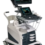
Product Overview
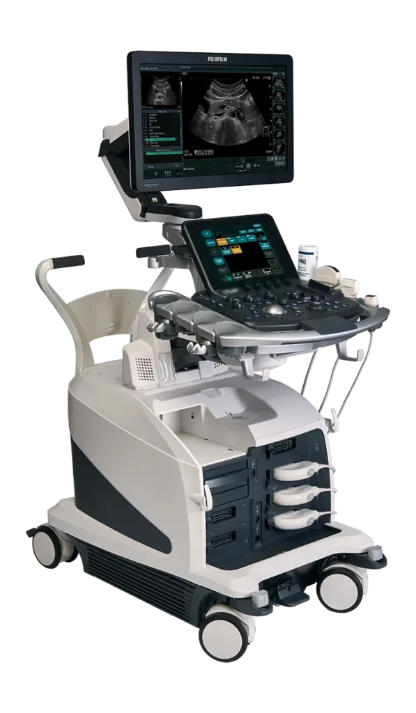
The Arietta 750 DeepInsight uses Fujifilm’s Variable Beamformer, which is designed to optimize both the transmission and the reception of ultrasound waves, resulting in clearer images. The machine also uses DeepInsight technology to provide improved clarity even in larger patients.
Top features of the Fujifilm Arietta 750DI include the following:
- DeepInsight technology extracts meaningful information from scans and reduces noise.
- Advanced probes make the scan experience both comfortable and effective for patients and providers.
- Technology such as detective flow imaging and carving imaging helps your team gain more insight from every exam.
- A compact footprint means this powerful machine can fit into tight exam spaces.
Core Features of the Airetta 750 DeepInsight
The Arietta DI produces high-quality images through its combination of innovative technology and optimized radiation. Scans take less time and offer more information, resulting in a better experience for both your patients and your staff.
Core features include the following:
- High-quality imaging capabilities
- DeepInsight technology
- Innovative transducers
Ultrasound exams should provide medical teams with significant information about patient health. Legacy machines are unwieldy, and they often produce blurry, fuzzy images. The Airetta 750DI is much more powerful, and it’s capable of delivering crisp images for patients of all sizes.
Fujifilm’s proprietary DeepInsight technology improves image quality, even in larger patients. Machine learning allows this machine to recognize anatomical landmarks and surface important information while reducing noise and blurring.
Images move through DeepInsight automatically, with no operator programming required. A natural representation of the tissue structure emerges, providing just what your operators need.
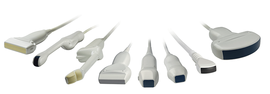
Every patient and exam is different. The Airetta 750DI ensures your team can respond to all diagnostic demands. Several types of transducers are available, including versions for the abdomen, vascular tissues, thyroid, and more.
Each transducer has a different frequency range and scan width. Operators can switch machines quickly until they find the version that’s right for their exam.
Technology
The Airetta 750DI includes technology that can improve workflows and efficiency. The machine is a pleasure for your technicians, as it’s ergonomically friendly and capable of delivering high-quality images very quickly.
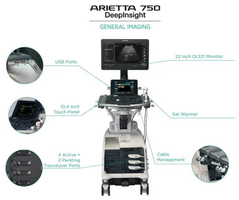
The Airetta 750DI’s control panel is height-adjustable and capable of moving from 48 inches to the floor. Your team can move the controls instead of stretching and straining during lower extremity exams.
The machine’s monitor is mounted on an articulated arm. Your team members can push and pull the screen as needed, rather than stretching to see their work.
The machine includes preset configurations, allowing your staff to press a button and start an exam without extensive programming. Automated image optimization controls ensure they can focus on patient care instead of image settings.
Ultrasound exams often involve routine steps and settings. A preprogrammed assistant handles these repetitive steps, allowing exams to move faster and reducing operator fatigue and frustration.
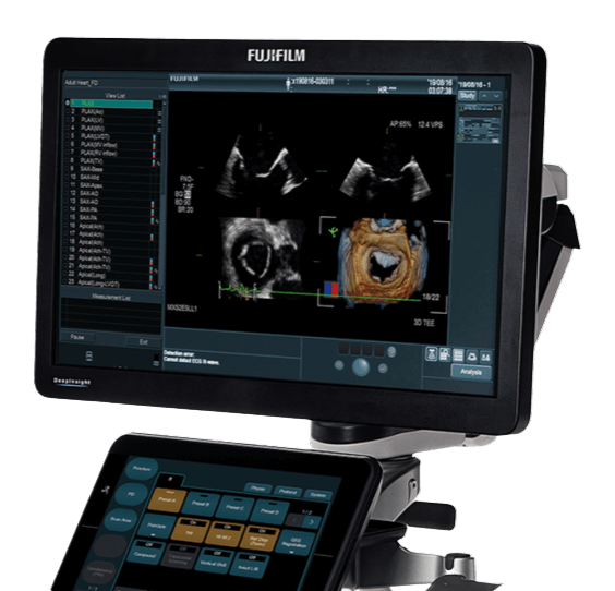
The Arietta 750DI has a 22-inch OLED monitor that’s crisp, bright, and easy to see. This monitor uses technology that improves contrast resolution, so even small details are easier to view. The monitor also reduces angle dependency, so images look good from any perspective.
The Airetta 750DI provides high-quality images that your entire diagnostic team will value. The machine is particularly adept at screening tissues within the breasts, gastrointestinal tract, kidneys, and pancreas.
Contact Us Today to Learn More
At Medical Outfitters, we can help you understand how the Fujifilm Arietta 750 DeepInsight works and what sets it apart from other ultrasound machines.
We have staff members available to walk you through the sales process, and we can perform routine maintenance to ensure your investment pays off in the long term. Contact us to get started today.
Request a Quote Today
Get in touch with Medical Outfitters today to request your personalized quote and outfit your facility with confidence.

