
Reviewed 23 September 2026. Facts, specifications and regulatory references on this page were checked against primary sources.
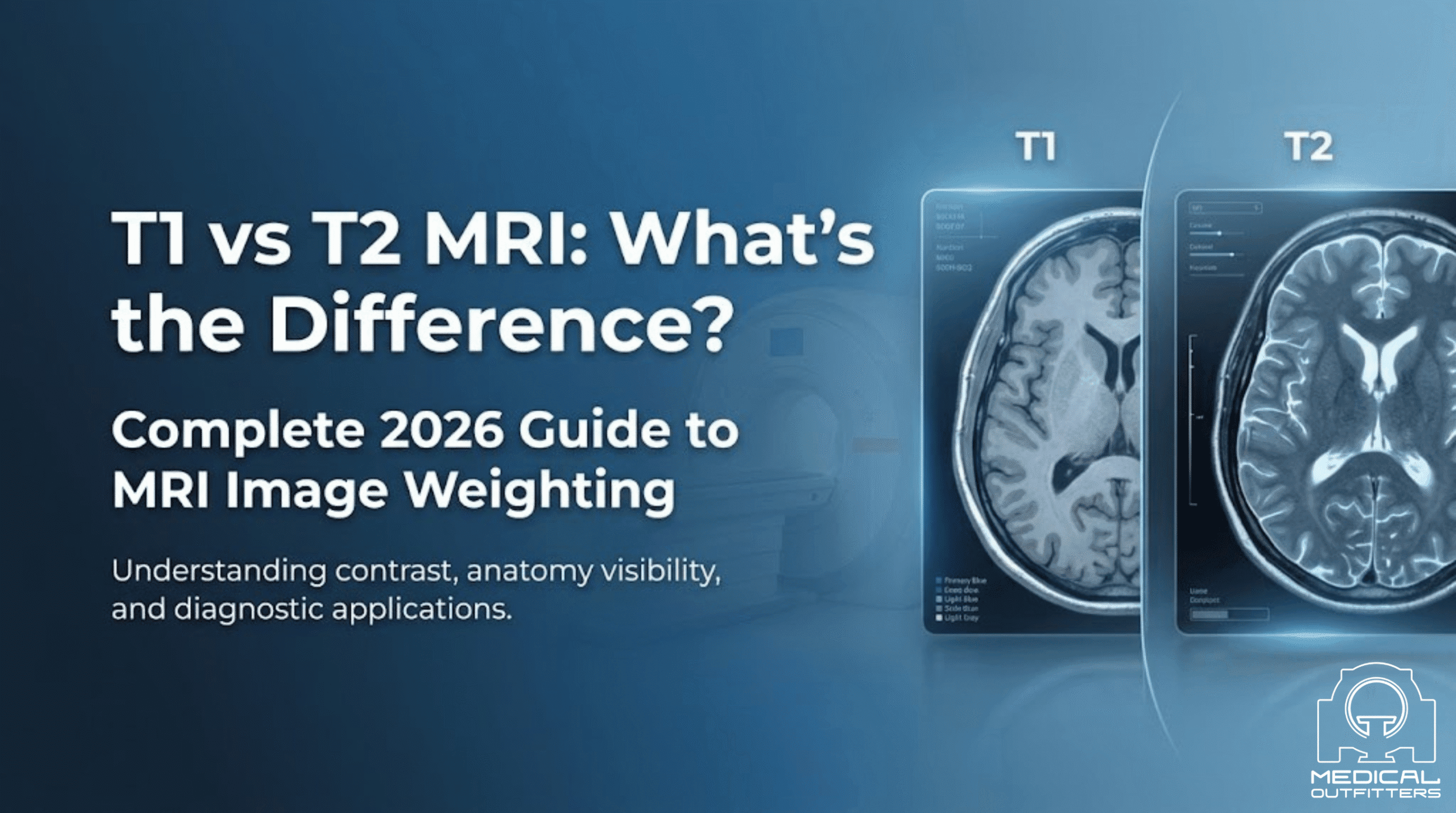
T1 vs T2 MRI is the first distinction anyone working in medical imaging has to get straight, from radiologists and technologists to referring physicians making diagnostic decisions. The two sequences answer different questions. These two signal weighting types form the backbone of clinical MRI interpretation, yet the distinction between them causes confusion even among experienced practitioners.
This guide explains what T1 and T2 MRI are, how each sequence works, and what tissues look bright or dark on each. It also covers when your clinical situation calls for one over the other. People search this as T1 versus T2 MRI, MRI T1 vs T2 and T2 weighted MRI; they are the same question, updated here for 2026.
T1 vs T2 MRI at a Glance
- T1-weighted images use a short repetition time (about 300 to 500 ms) and a short echo time (under 30 ms); fat and gadolinium-enhanced tissue read bright, free water dark. Source: Radiology Cafe, FRCR physics notes
- T2-weighted images use a long repetition time (over 1,500 ms) and a long echo time (over 90 ms); free water, edema and most pathology read bright. Source: Radiology Cafe, FRCR physics notes
- Proton density weighting uses a long repetition time with a short echo time, minimizing both T1 and T2 effects. Source: Radiology Cafe
- MRI produces its images with a magnetic field and radio waves and involves no ionizing radiation. Source: US FDA
Understanding MRI Signal Weighting
MRI does not produce a single type of image. Instead, the system can be programmed to emphasize different tissue properties by adjusting timing parameters, specifically the repetition time (TR) and echo time (TE). These adjustments produce “weighted” images that make certain tissues appear bright or dark depending on their physical characteristics.
The two dominant forms of weighting in clinical practice are T1 weighting and T2 weighting. A third, proton density (PD) weighting, exists but is less commonly used. Understanding T1 and T2 is the key to reading the majority of MRI studies.
T1-weighted MRI uses a short TR and short TE. In T1-weighted images, signal intensity reflects how quickly hydrogen protons in tissue return to their resting state after being disturbed by the MRI radiofrequency pulse, a process called longitudinal (T1) relaxation.
What Appears Bright on T1?
Tissues with short T1 relaxation times recover quickly and therefore appear bright (hyperintense) on T1-weighted images:
- Fat: adipose tissue, bone marrow, and subcutaneous fat all appear very bright
- Gadolinium contrast: intravenous contrast agents dramatically shorten T1, causing enhanced areas to appear bright white
- Protein-rich fluids: mucus, certain cysts, and proteinaceous collections
- Subacute hemorrhage (methemoglobin): blood at a specific stage of breakdown appears bright
- Melanin: relevant in melanoma imaging
What Appears Dark on T1?
- Water and simple fluids: cerebrospinal fluid (CSF), edema, cysts
- Cortical bone: almost no mobile protons
- Air and calcification
- Most pathology: tumors, inflammation, and infarcts typically appear dark or hypointense
When Is T1 MRI Used?
T1-weighted sequences are the workhorse for:
- Post-contrast imaging: identifying breakdown of the blood-brain barrier, tumor vascularity, and inflammatory enhancement
- Anatomical detail: excellent contrast between fat, soft tissue, and bone
- Liver imaging: characterizing hepatic lesions, detecting fat infiltration
- Musculoskeletal studies: bone marrow assessment, joint anatomy
- Detecting fat: lipomas, teratomas, adrenal myelolipoma
- Assessing subacute hemorrhage
T2-weighted MRI uses a long TR and long TE. Rather than measuring how quickly tissue recovers, T2 weighting measures how quickly hydrogen protons lose phase coherence with each other, called transverse (T2) relaxation.
What Appears Bright on T2?
Tissues with long T2 relaxation times retain their signal longer and appear bright (hyperintense) on T2-weighted images:
- Free water and simple fluids: CSF, urine, bile, edema all appear very bright
- Most pathology: tumors, inflammation, infection, infarction, demyelination all have elevated water content and appear bright
- Edema: T2 is exquisitely sensitive to any process that increases tissue water
- Joint fluid and effusions: bright fluid outlines the intermediate-signal hyaline cartilage, important in joint imaging
- Nucleus pulposus of healthy intervertebral discs
What Appears Dark on T2?
- Cortical bone and calcification
- Fibrous tissue: tendons, ligaments, fibrocartilage
- Air
- Dehydrated nucleus pulposus: a reliable marker of disc degeneration
- Hemosiderin and chronic hemorrhage: appears very dark, and blooms on gradient echo, T2* and SWI sequences
- Melanin in some contexts
When Is T2 MRI Used?
T2-weighted sequences are preferred for:
- Detecting pathology: because most diseases increase tissue water, T2 is the most sensitive sequence for finding abnormalities
- Brain and spine imaging: MS plaques, strokes, tumors, and infection all show up clearly
- Pelvic imaging: exquisite soft tissue contrast for prostate, uterus, and ovaries
- Cardiac MRI: edema assessment (fibrosis and scar are assessed with T1-based late gadolinium enhancement)
- Abdominal imaging: liver lesion characterization, biliary tree assessment
- Musculoskeletal: ligament and cartilage injury, bone marrow edema
T1 vs T2 MRI: Side-by-Side Comparison
| Feature | T1-Weighted | T2-Weighted |
|---|---|---|
| TR (Repetition Time) | Short (~400-600 ms) | Long (~2,000-5,000 ms) |
| TE (Echo Time) | Short (~10-20 ms) | Long (~80-120 ms) |
| Fat appearance | Bright (hyperintense) | Intermediate to slightly bright |
| Free water / CSF | Dark (hypointense) | Bright (hyperintense) |
| Most pathology | Dark | Bright |
| Gadolinium contrast | Dramatically brightens | Less affected |
| Best for | Anatomy, post-contrast, fat | Pathology detection, fluid |
| Scan time | Faster | Longer (fast spin echo shortens it considerably) |
The Simple Memory Rule
Radiologists and technologists often use this practical mnemonic:
T1 = “White Fat, Dark Water”
T2 = “White Water, Fat Stays Bright (unless suppressed)”
On T1: fat is white, water is dark.
On T2: water is white, and fat is intermediate to bright on the fast spin echo sequences used today, which is why fat suppression is added when fat needs to be dark.
CSF (pure water) is the clearest example: on T1 it appears black; on T2 it appears bright white. Use the CSF in any brain scan as your calibration reference.
T1 vs T2 in Common Clinical Scenarios
A complete brain MRI for tumor staging will include both T1 and T2:
- T2/FLAIR shows the extent of tumor infiltration and surrounding edema, which typically extends beyond the contrast-enhancing core
- T1 with gadolinium shows breakdown of the blood-brain barrier, identifying the active, high-grade component
Neither sequence alone is complete. The combination defines both tumor biology and extent.
MS plaques are best seen on T2 and FLAIR as hyperintense (bright) white matter lesions. Active plaques showing current inflammation will additionally enhance on T1 with gadolinium: distinguishing new from old disease and guiding treatment decisions.
- T2 shows healthy discs as bright (high nucleus pulposus water content) and identifies degenerated “black discs” that have lost hydration
- T2 also reveals spinal cord edema, compression, and nerve root compression
- T1 with contrast identifies post-surgical fibrosis vs. recurrent disc herniation and detects leptomeningeal disease
Liver MRI uses a complex multi-phase protocol, but the core principles apply:
- T1 in-phase and out-of-phase detects fatty infiltration (signal dropout on out-of-phase T1)
- T2 characterizes lesions: hemangiomas are very bright; hepatocellular carcinoma is moderately bright; simple cysts are very bright; metastases are moderately bright
- Dynamic T1 with gadolinium (arterial, portal venous, and delayed phases) characterizes enhancement patterns
- T2 with fat suppression is the most sensitive sequence for bone marrow edema, muscle injury, ligament tears, and joint effusions
- T1 provides anatomy, bone marrow signal assessment, and detection of fatty marrow replacement
FLAIR, Advanced Sequences & System Quality
In brain imaging you will frequently encounter FLAIR (Fluid Attenuated Inversion Recovery): a modified T2-weighted sequence that suppresses the bright CSF signal. This is critically important because:
- Periventricular lesions (MS plaques, subtle infarcts) can be hidden by bright adjacent CSF on standard T2
- FLAIR removes this masking, revealing pathology adjacent to fluid spaces
- FLAIR has become the standard sequence for detecting white matter disease, cortical lesions, and subarachnoid pathology
For completeness: proton density weighting uses a long TR and short TE, minimizing both T1 and T2 effects. PD images reflect the density of hydrogen protons in tissue, providing excellent contrast for cartilage evaluation in joints. PD fat-saturated sequences have largely replaced traditional T2 for knee and shoulder imaging in many protocols.
Modern MRI systems offer dozens of specialized sequences built on T1 and T2 principles:
- DWI (Diffusion-Weighted Imaging): detects acute stroke within minutes, also characterizes tumors and abscesses. Restricted diffusion appears bright on DWI and dark on ADC maps.
- SWI (Susceptibility-Weighted Imaging): extremely sensitive to blood products, calcification, and iron, a highly sensitive T2*-based technique
- MR Spectroscopy: measures metabolite concentrations within a defined voxel
- BOLD fMRI: functional mapping based on T2* changes during brain activity
- MR Elastography: assesses tissue stiffness, used in liver fibrosis staging
These advanced sequences supplement, but do not replace, fundamental T1 and T2 interpretation.
The clarity and diagnostic value of T1 and T2 images depends heavily on field strength and coil selection.
Field Strength
- 1.5 Tesla systems are the clinical standard and provide excellent T1 and T2 images for most indications
- 3.0 Tesla systems offer higher signal-to-noise ratio, allowing thinner slices, faster scans, and better spatial resolution, particularly valuable for brain, spine, and musculoskeletal imaging
- Open MRI systems (typically 0.25T to 1.2T) provide patient-friendly scanning but with lower signal, acceptable for many routine studies but limited for demanding applications
Coil Selection
Phased-array receiver coils dramatically improve signal-to-noise. Surface coils positioned close to the anatomy of interest provide superior image quality compared to the body coil.
Protocol Optimization
Standard T1 and T2 sequences are just starting points. Experienced radiologists and technologists optimize TR, TE, flip angle, slice thickness, and matrix size based on the clinical question. Current-generation systems add AI at the acquisition and reconstruction stage instead: Fujifilm’s Synergy DLR deep learning reconstruction with AutoPose and AutoExam workflow automation, and Esaote’s e-SPADES HyperSpeed and HyperClarity, shorten scans and recover image quality, but the protocol parameters themselves are still set by the team.
Choosing the Right MRI System for Your Practice
If you are evaluating MRI systems for purchase, the ability to deliver high-quality T1 and T2 images across your patient population is the fundamental performance standard. Key considerations in 2026:
1.5T systems with modern gradient performance (≥30 mT/m amplitude, ≥150 mT/m/ms slew rate) deliver excellent T1 and T2 quality for most clinical applications. Fujifilm Healthcare’s ECHELON Smart (1.5T) and Oasis Velocity (1.2T open) and Esaote’s open MRI platforms are strong options for facilities serving diverse patient populations including larger patients.
3T systems offer meaningful advantages in T2-weighted sequence resolution and speed. The incremental cost must be weighed against patient volume and case mix.
Open and compact MRI designs have improved significantly. Esaote’s G-Scan Brio (weight-bearing, 0.25T) and O-Scan (dedicated extremity, 0.31T) provide excellent T1/T2 quality for their intended anatomical focus.
Facilities should evaluate total cost of ownership including service contracts, helium consumption (zero-helium and helium-free systems are now clinically available), and vendor support infrastructure.
T1 vs T2 MRI: Where to Go Next
The sequence is only half the decision. The scanner that produces it decides how fast you get the images and how much detail they carry.
- Open MRI vs closed MRI Which configuration suits your caseload, and what field strength costs you
- Wide bore MRI systems A 70 cm opening at full 1.5T or 3T, and whether the extra room costs image quality
- MRI machine cost and price guide System price ranges by field strength and condition
- What an MRI quench is Why superconducting systems need cryogens and a vent
- Our MRI inventory Open, wide bore and standard bore systems currently available
For background on the imaging physics itself, the FDA overview of MRI is a reliable starting point. If you are weighing a purchase rather than reading a scan, tell us what you are trying to image and we will tell you which platform actually covers it.
For the difference between the two modalities rather than two sequences, see our MRI vs CT comparison.
T1 vs T2 MRI: Common Questions Answered
T2 and FLAIR are generally more sensitive for detecting tumor extent and surrounding edema. T1 with gadolinium contrast is superior for characterizing active enhancement, assessing blood-brain barrier breakdown, and evaluating vascularity. A complete tumor evaluation requires both.
Fat has a very short T1 relaxation time (it recovers quickly), giving it high signal on T1-weighted sequences. Fat has a relatively short T2 relaxation time as well, which would make it dark on conventional T2 spin echo, though in practice fat appears intermediate to bright on the fast spin echo T2 sequences used clinically, because the closely spaced refocusing pulses suppress J-coupling signal loss. Fat-saturation techniques are used to specifically suppress the fat signal when needed.
Hyperintense on T2 means the tissue appears brighter than surrounding tissue on T2-weighted images, indicating high water content. This is the appearance of free fluid (CSF, effusion), edema and most pathology (tumors, infection, infarction). Healthy hyaline cartilage is intermediate rather than bright.
Yes. T1 and T2 are intrinsic tissue properties; they exist without any contrast agent. Contrast (typically gadolinium-based) modifies the T1 signal of tissues it accumulates in, but the underlying T1 and T2 sequences are performed regardless. Many diagnostic MRI protocols are performed entirely without contrast.
Modern sequences using parallel imaging and compressed sensing can acquire T2-weighted brain scans in under 2 minutes. Standard T1 post-contrast sequences run 3-6 minutes. Full brain MRI protocols including multiple T1 and T2 sequences typically complete in 20-45 minutes depending on complexity.
Hypointense, or decreased signal, means the tissue returns less signal than the tissue around it on that particular sequence, and it has to be read against the weighting. On T1-weighted images low signal is normal for fluid and for most water-rich pathology such as edema, cysts and many tumors. On T2-weighted images low signal points the other way, toward fibrous tissue, calcification, hemosiderin from old bleeding, dense cortical bone or air. The same lesion can be dark on one sequence and bright on the other, which is exactly why radiologists read both.
Dark on T1 and bright on T2 is the most common pattern in MRI and is the signature of increased water content. Edema, inflammation, infection, many tumors and simple cysts all behave this way. It says a lesion is water-rich; it does not say what the lesion is. Contrast enhancement, diffusion imaging, the shape and location of the lesion and the clinical history are what narrow it down.
STIR is a T2-based sequence that suppresses the signal from fat, so anything bright on STIR is a water-rich process that would otherwise be hidden inside fat-containing tissue such as bone marrow or subcutaneous fat. Bone marrow edema after a fracture or stress injury, muscle injury, soft-tissue inflammation and infection are the usual causes. STIR is also useful when gadolinium is not being given.
Small bright spots on T2 or FLAIR images, most often in the white matter of the brain. A few are common with age and with small-vessel disease linked to hypertension and diabetes; more numerous or unusually placed foci can reflect demyelination as in multiple sclerosis, migraine, prior infection or other processes. Number, size, location and the patient history decide the meaning, which is why a radiologist reports them in context rather than as a diagnosis on their own.
A lesion that is dark on both T1 and T2 in a vertebral body usually contains little water: sclerotic or dense bone, fibrosis, or heavy mineralization. Osteoblastic, bone-forming metastases, bone islands and the sclerotic stage of endplate degenerative change are typical examples, and old fractures with dense healing can look the same. Because bone-forming tumors sit in this group, a new dark-on-both lesion is followed up with the history, CT and sometimes a bone scan.
Key Takeaways
T1 and T2 MRI represent the two fundamental tools of magnetic resonance imaging:
- T1: Short TR/TE, fat is bright, water is dark, ideal for anatomy, fat detection, and post-contrast imaging
- T2: Long TR/TE, water is bright, fat is intermediate, ideal for pathology detection and fluid characterization
The mnemonic is simple: T1 = white fat, T2 = white water. In clinical practice, most MRI protocols include both because they answer different diagnostic questions: T2 finds the abnormality, T1 with contrast characterizes it.
Understanding which sequence you are looking at, and what the signal patterns mean, is the foundation of MRI interpretation regardless of whether you are reviewing a brain scan, a shoulder study, or a whole-body oncology protocol.
Medical Outfitters provides new, certified pre-owned, and refurbished MRI systems including Fujifilm Healthcare Echelon series, Esaote G-Scan, and Esaote O-Scan dedicated extremity MRI. Our team supports facilities across the United States, Caribbean, and Latin America with installation, rigging, cryogenic services, and ongoing clinical support.
