A CT scan with contrast uses an iodine-based dye, injected into a vein or swallowed, to make blood vessels, organs and abnormal tissue stand out from everything around them. A CT scan without contrast images the body as it is: bone, lung, calcium, stones, bleeding and gross anatomy show clearly, but a tumor, an infection or a blocked artery can hide in tissue of the same density. The radiologist chooses between the two, or orders both, based on the question the scan has to answer. For the facility that owns the scanner, contrast is also a workflow and an equipment decision: the injector, the bolus-tracking software, the generator and tube, and dual-energy imaging all exist to get more from each dose of dye.
This guide explains what CT contrast is, what a CT with contrast shows that a CT without contrast cannot, which exams use which, how a contrast scan runs from screening to discharge, the side effects and the safety rules in the current ACR Manual on Contrast Media, and what contrast means when a hospital or imaging center is choosing a CT scanner. Medical Outfitters sells, installs and services CT scanners across the United States, Puerto Rico and the Caribbean, so the last part is written for the people who will run the room.
- Without contrast: bone, lung, kidney stones, acute bleeding in the head, calcium scoring, low-dose lung screening and most follow-up of known findings.
- With IV contrast: blood vessels (CT angiography), solid organs, tumors, infection and abscess, bowel and lymph nodes, trauma surveys and most cancer staging.
- With and without: two passes through the same anatomy, used when the change between them is the finding, such as a kidney or adrenal mass or a head scan for a possible tumor.
- Safety: acute reactions to modern non-ionic contrast run at roughly 0.2 to 0.7 percent and serious reactions at about four in 10,000; kidney function and allergy history decide who is screened more carefully.
- For the scanner: a power injector, bolus tracking, a generator and tube that can hold an arterial phase, and increasingly dual-energy acquisition.
What Is Contrast in a CT Scan?
CT measures how much X-ray each small volume of tissue absorbs and reports it in Hounsfield units: water is 0, air is about minus 1,000, dense bone runs from several hundred to more than 1,000. Soft tissues cluster in a narrow band between about 20 and 80, which is the problem. A liver tumor, the liver around it, a loop of bowel and the artery feeding all three sit within a few Hounsfield units of one another, and on a plain scan they can look the same.
CT contrast, often called contrast dye, is an iodine-based liquid that absorbs far more X-ray than tissue. When it is injected into a vein it travels with the blood, so for a minute or two the arteries, then the veins, then every organ with a blood supply light up in proportion to how much blood they receive. A tumor with its own blood vessels enhances differently from the tissue around it; an abscess has a bright rim and a dark center; a blocked artery simply stops being bright. The dye is cleared by the kidneys over the following hours.
The agents used today are non-ionic, low-osmolar or iso-osmolar iodinated contrast media. They replaced the older ionic, high-osmolar agents in the 1990s and cut the reaction rate several-fold. Three routes are common:
Intravenous (IV) contrast. Injected through a cannula, usually in the arm, by a power injector at a set rate. This is what people mean by a CT scan with contrast.
Oral contrast. A diluted iodine or barium drink taken over an hour or so before an abdominal scan so the bowel is outlined. Many departments now use neutral oral contrast (water or a low-density preparation) for CT enterography and for scans where bright bowel would hide the finding.
Rectal, intrathecal and other routes. Less common, used for specific questions such as a suspected bowel leak or a myelogram.
CT contrast is not the same substance as MRI contrast, which is gadolinium based and works by changing how tissue responds to the magnetic field. Our MRI vs CT scan guide covers that difference and when each modality is chosen.
CT Scan With Contrast vs Without: What Each One Shows
The two scans are not better and worse versions of each other. A non-contrast CT is the right exam for some questions and the wrong one for others, and the same is true of a contrast study.
What a CT scan without contrast shows well
Anything whose density is naturally far from soft tissue. Fractures and bone lesions. Lung nodules, pneumonia, emphysema and the low-dose lung cancer screening scan. Kidney stones and most ureteric stones, which are the classic reason for a non-contrast CT of the abdomen. Fresh bleeding in the head, which is why the first scan after a stroke or a fall is almost always without contrast. Coronary artery calcium scoring. Sinuses, temporal bones and the spine. Follow-up of a known finding whose size is the only question.
What a CT scan with contrast adds
Everything that depends on blood supply or on telling one soft tissue from another. CT angiography of the head, neck, chest, abdomen or legs, including the pulmonary CT angiogram for a suspected clot. Liver, pancreas, kidney and adrenal masses, where the way a lesion enhances in each phase is the diagnosis. Staging of most cancers, where contrast separates lymph nodes from vessels and shows metastases in the liver. Infection and abscess. Bowel wall inflammation, ischaemia and perforation. Trauma surveys of the chest, abdomen and pelvis, where active bleeding shows as contrast leaking out of a vessel.
Why a doctor orders a CT with and without contrast
When the change between the two is the finding. A kidney mass that measures 20 Hounsfield units higher after contrast is enhancing and therefore solid; one that does not change is a cyst. Adrenal lesions are characterized by how fast they wash the contrast out again. A head CT for a possible tumor or infection is often done both ways. The cost of the second pass is extra radiation dose and time, which is why the protocol is chosen per question rather than applied to everyone.
Which CT Exams Use Contrast and Which Do Not
| Exam | Usual protocol | Why |
|---|---|---|
| Head CT, acute (stroke, trauma, headache) | Without contrast | Fresh blood and skull fractures show without dye; contrast would mask bleeding |
| Head CT for tumor, infection or metastases | With contrast, often with and without | Enhancement pattern separates tumor, abscess and normal brain |
| CT angiography (head, neck, chest, aorta, legs) | With IV contrast, timed to the arterial phase | The vessels are the subject; without dye they are invisible |
| Chest CT for nodules or lung disease | Without contrast | Air and lung tissue give natural contrast |
| Chest CT for cancer staging, infection or lymph nodes | With IV contrast | Separates nodes, vessels and masses in the mediastinum |
| CT pulmonary angiogram (clot) | With IV contrast, pulmonary arterial phase | A clot appears as a filling defect in a bright artery |
| Abdomen and pelvis, kidney stones | Without contrast | Stones are dense; dye in the urine would hide them |
| Abdomen and pelvis, pain, infection, cancer | With IV contrast, portal venous phase; oral contrast by department policy | Organs, bowel and nodes separate from one another |
| Liver, pancreas, kidney mass characterisation | Multiphase: without, arterial, portal venous and sometimes delayed | The enhancement curve of the lesion is the diagnosis |
| Trauma survey | With IV contrast | Active bleeding and organ injury show as contrast extravasation |
| Coronary calcium score | Without contrast | Calcium is the subject |
| Coronary CT angiography | With IV contrast, ECG-gated | The arteries must be bright and still |
| Spine, sinuses, temporal bone, extremities | Without contrast | Bone detail; contrast only when infection or tumor is suspected |
| Low-dose lung cancer screening | Without contrast | Screening of healthy people at the lowest dose |
Protocols vary between departments and radiologists, and the referring question always wins. The table describes the common practice, not a rule.
How a CT Scan With Contrast Is Done
Screening. Before any IV contrast the technologist confirms the patient’s kidney function, allergy history, prior contrast reactions, pregnancy status and current medicines. The ACR Manual on Contrast Media is the reference most United States departments follow for who needs a recent creatinine or eGFR, who needs premedication and who should be imaged another way.
IV access and the injector. A cannula, usually 18 to 22 gauge in the forearm or antecubital vein, is connected to a power injector. The injector delivers a set volume of contrast at a set rate, commonly 60 to 150 mL of contrast at 2 to 5 mL per second for a general adult study, followed by a saline chaser that pushes the dye through and reduces the dose needed. Dual-head injectors hold the contrast and the saline in separate syringes; some departments use multi-dose delivery systems to cut waste.
A non-contrast pass, if ordered. For a with-and-without protocol the plain scan runs first.
Injection and timing. The scan has to catch the dye in the right place. Bolus tracking places a region of interest on the aorta and starts the scan automatically when the Hounsfield value crosses a threshold; a test bolus does the same job with a small injection first. The arterial phase comes about 20 to 35 seconds after injection starts, the portal venous phase at 60 to 80 seconds, and delayed phases from three to fifteen minutes depending on the organ.
The scan itself. Each phase is a few seconds on a modern 64-slice or faster scanner. The patient feels a warm flush spreading through the body, sometimes a metallic taste, and often the sensation of having passed urine, which they have not. All three fade within a minute or two.
Observation and discharge. Most reactions that will happen do so within the first twenty minutes, so the cannula stays in and the patient is observed before leaving. Drinking normally over the rest of the day helps the kidneys clear the contrast, which is largely gone within 24 hours in someone with normal kidney function.
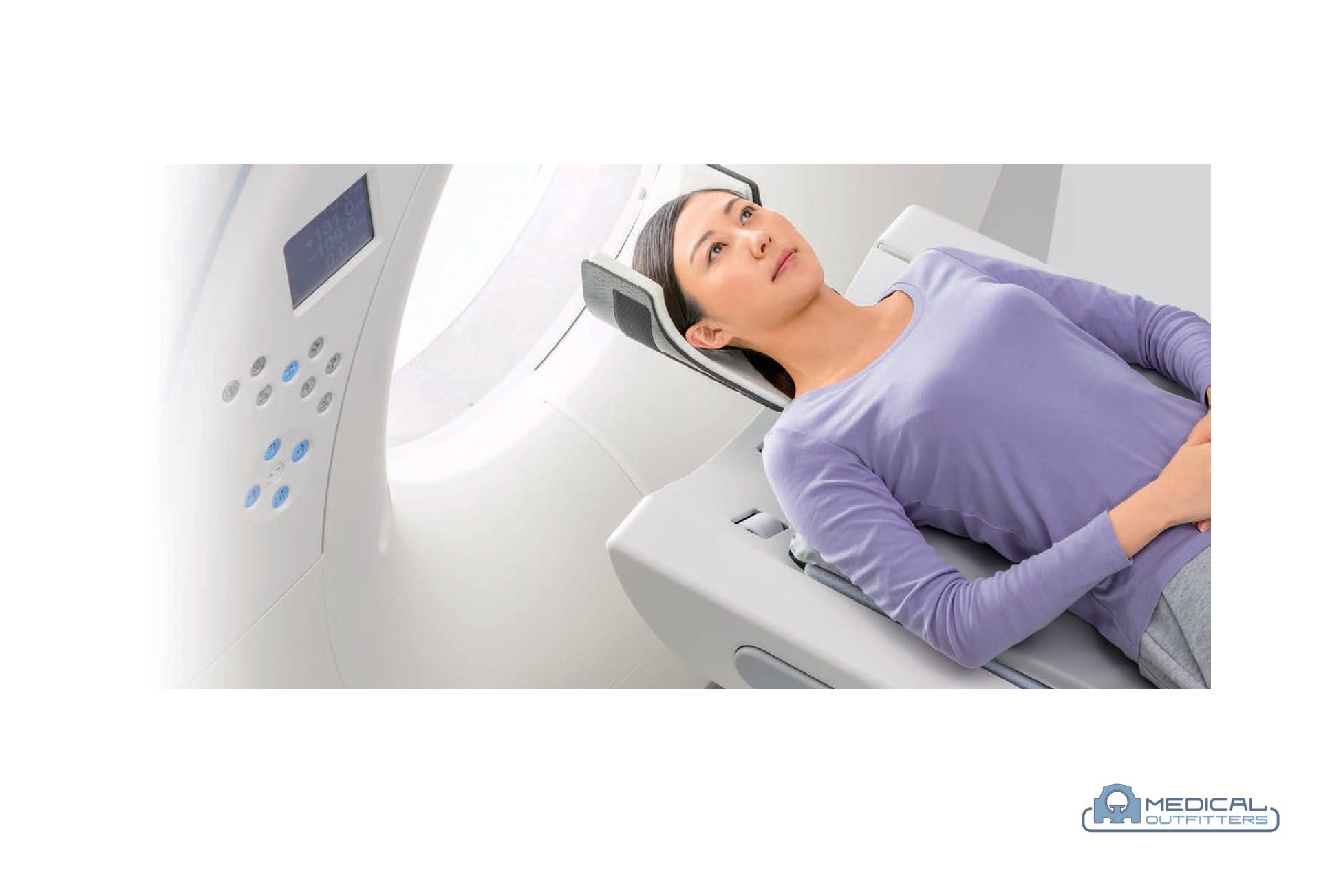
CT Contrast Side Effects and Who Needs Extra Care
Expected sensations. Warmth, flushing, a metallic taste and nausea are physiological responses to the injection, not allergic reactions. They are common and pass on their own.
Acute reactions. The ACR puts the overall rate of acute adverse reactions to modern non-ionic, low-osmolar contrast at roughly 0.2 to 0.7 percent of injections, the large majority of them mild: hives, itching, mild nasal congestion. Serious reactions, meaning bronchospasm, significant swelling or a drop in blood pressure that needs treatment, are rare at about four in 10,000. Every CT department that injects contrast keeps an emergency kit, trains its staff in reaction management and has a physician available.
Who is at higher risk of an allergic-like reaction. People with a prior reaction to iodinated contrast, who are at several times the baseline risk, and people with significant asthma or multiple allergies. A shellfish or iodine allergy is not a contrast allergy and is not a reason to withhold the exam; the ACR has said so for years. For patients with a prior moderate or severe reaction the options are premedication with corticosteroids and an antihistamine on the ACR schedule, a different agent, or a different test.
Kidneys. Contrast-associated acute kidney injury is far less common than it was once believed to be, and the ACR’s current position is that an eGFR below 30 mL/min/1.73 m², or an acute kidney injury, is the threshold at which IV iodinated contrast becomes a relative contraindication to be weighed against the benefit of the scan. Patients above that threshold with stable kidney function do not need special preparation. Patients on dialysis can receive contrast with their next session scheduled normally.
Metformin. The 2024 ACR guidance is that patients with an eGFR of 30 or above and no acute kidney injury do not need to stop metformin before or after IV contrast, and do not need a repeat kidney test afterwards. Patients below 30, or with an acute kidney injury, hold metformin at the time of the scan and for 48 hours and have their kidney function checked before restarting.
Pregnancy and breastfeeding. Iodinated contrast crosses the placenta, and no harm to the fetus has been documented from diagnostic doses; it is given in pregnancy when the scan is needed and the decision is shared with the patient. A breastfeeding mother can continue to feed normally: a tiny fraction of the dose reaches the milk and almost none of that is absorbed by the baby.
Thyroid. The iodine load is a consideration for patients with hyperthyroidism and for anyone scheduled for radioactive iodine treatment or a thyroid uptake scan, which should be delayed by several weeks after a contrast CT.
Extravasation. Occasionally the injected contrast leaks into the tissue around the cannula instead of the vein. Most cases cause swelling and discomfort that settle over a day or two; the injector’s pressure limit and the technologist’s check of the line before the main injection are the safeguards.
Oral Contrast: When the Drink Is Needed
Positive oral contrast, a dilute iodine or barium solution drunk over 60 to 90 minutes before the scan, makes the bowel bright so that a loop of intestine is not mistaken for a mass or an abscess. It is still used for suspected abscess, post-operative leaks and some oncology follow-up. Many departments have moved to neutral oral contrast, or to none at all, for general abdominal pain, trauma and CT enterography, because bright bowel contents hide the enhancing bowel wall that those exams are looking at. The choice is a department protocol decision and a throughput decision: an hour of drinking is an hour of waiting room.
What Contrast Means When You Buy a CT Scanner
Most of the volume in a general CT department is contrast-enhanced, and it is the contrast work, not the plain scans, that sets the equipment specification.
Speed and tube power. An arterial phase lasts a few seconds. The scanner has to cover the anatomy inside that window, which is why rotation time, detector coverage and the generator’s output matter more for CT angiography than for a sinus scan. Slice count is the headline number, and our guide to CT scanner types by slice count explains what 16, 64 and 128 slices change in practice.
Bolus tracking and protocol software. Automatic triggering, saved multiphase protocols and dose modulation are standard on current platforms but vary on older refurbished systems. Confirm the licenses are included and transfer with the scanner.
The injector. The power injector is bought or leased separately from the scanner, which is why our CT scanner cost guide lists it among the items quoted per project. A dual-head injector with saline chase, a contrast warmer and extravasation detection are the usual specification; integration with the scanner console so that the injection and the scan start together is worth asking about on any system.
Dual-energy and spectral CT. Scanning at two energies lets the software create a virtual non-contrast image from a contrast scan, which can replace the plain pass in a with-and-without protocol and save a phase of dose, and iodine maps that quantify enhancement. The Fujifilm SCENARIA View offers dual-energy acquisition; GE, Siemens, Philips and Canon each have their own implementation on their current platforms, which our comparison of CT scanner manufacturers covers.
Low-kV protocols. Scanning at 80 or 100 kV instead of 120 makes iodine brighter, so less contrast is needed for the same enhancement, which matters for patients with reduced kidney function and for the contrast budget. Lower kV asks more of the tube and relies on iterative reconstruction to control noise, which is a reason to buy a scanner with a documented tube and current reconstruction software rather than the oldest system that fits the room.
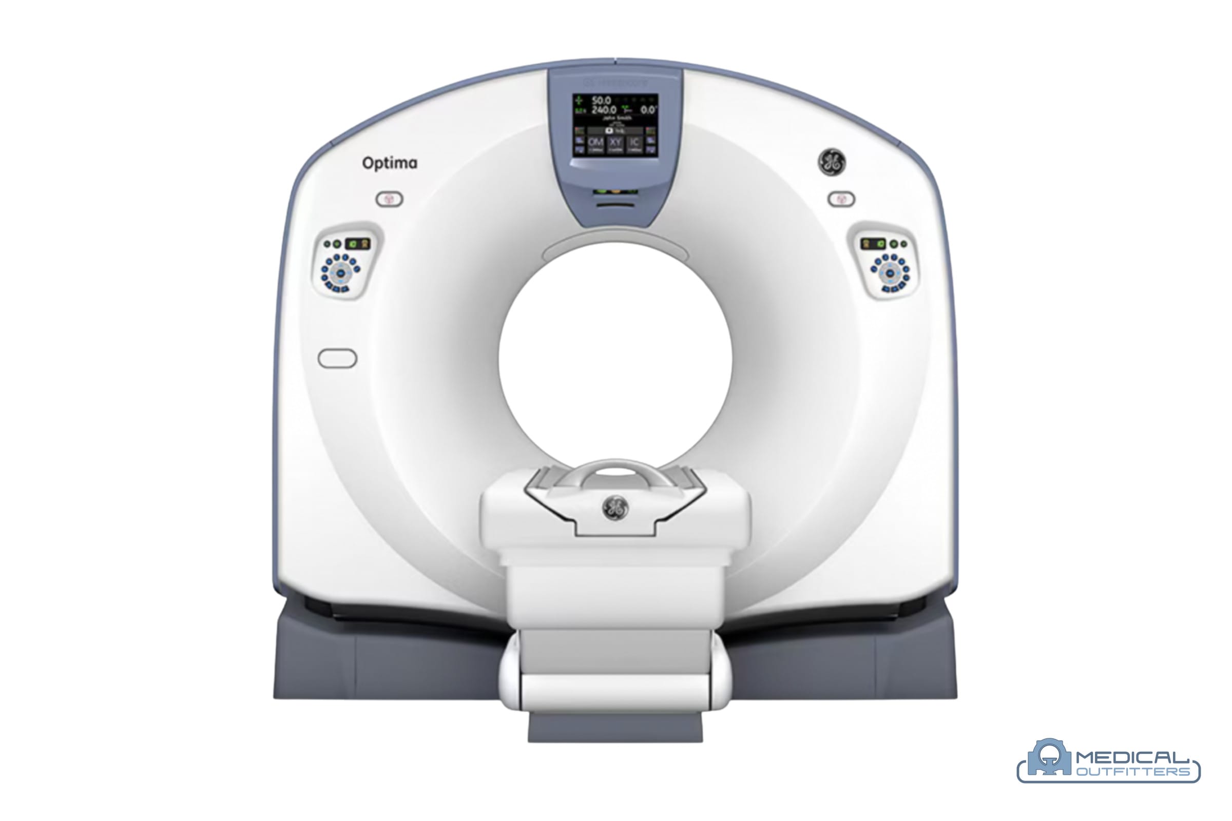
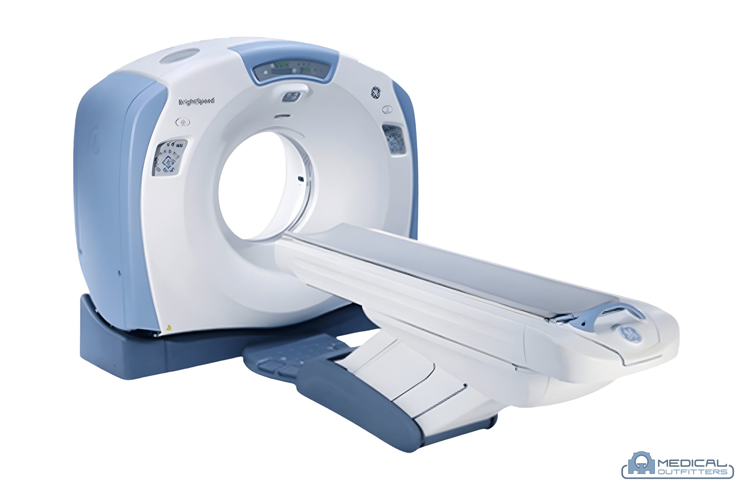
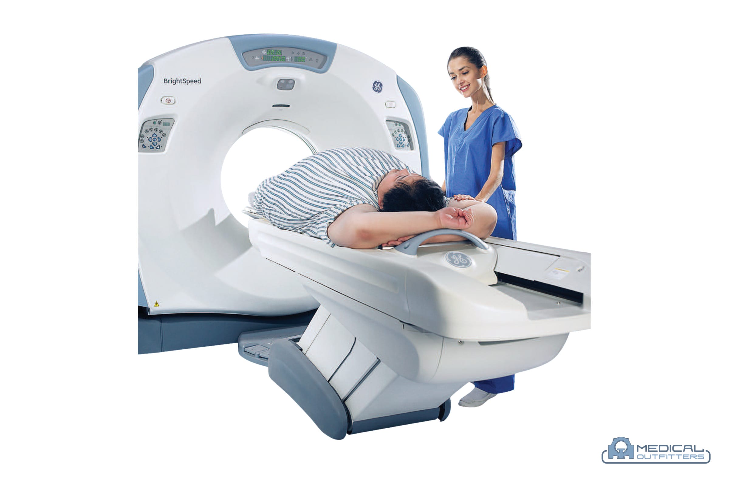
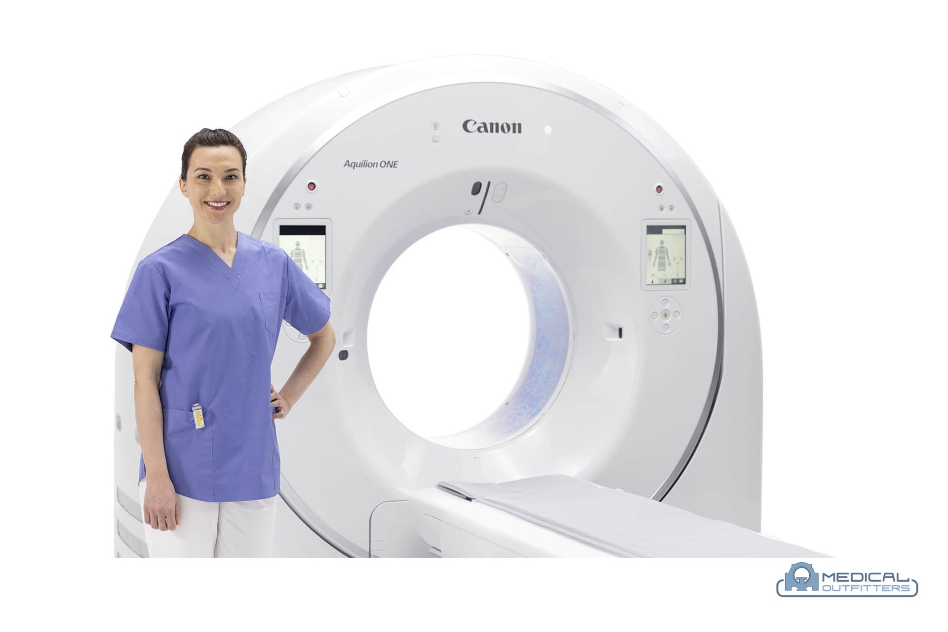
Refurbished options. A refurbished 64-slice system such as the GE Optima CT660 or the LightSpeed VCT handles routine contrast work, CT angiography included, at a fraction of the price of a new scanner. Cardiac CT angiography and the fastest trauma protocols are where the newer platforms, the GE Revolution family and the Canon Aquilion line among them, earn their premium.
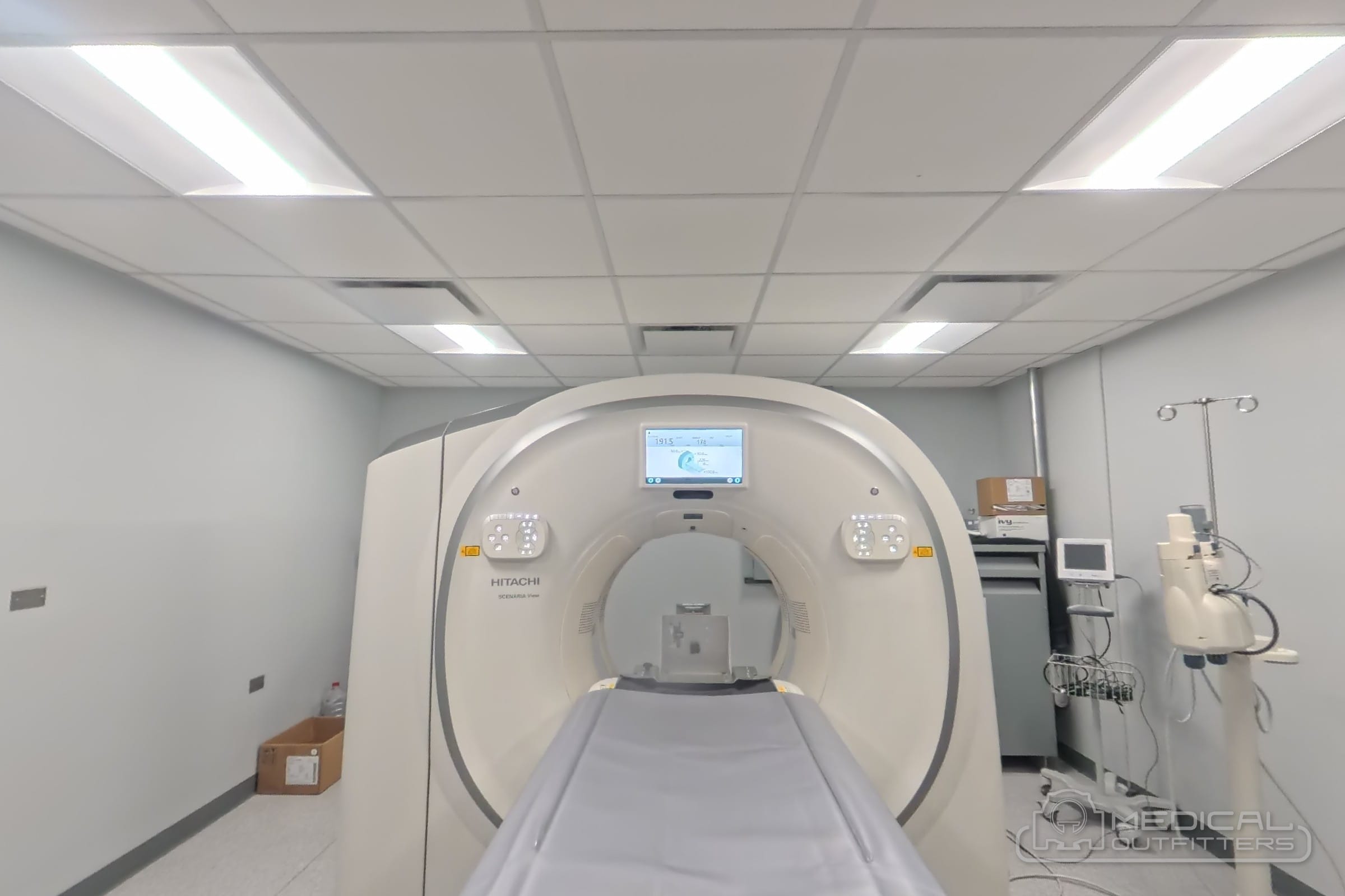
Medical Outfitters sells new Fujifilm CT scanners as an authorized dealer, including the Supria 128 and the SCENARIA View, and refurbished GE and Siemens systems through MO Select, delivered, installed and warrantied by our own engineers. Our CT scanner service team maintains the scanner, monitors the tube and keeps the protocol software current, and the full range of systems is on the CT scanners for sale page. To talk through a contrast workload and the scanner that fits it, contact us.
- CT scanner cost and price guide (2026)
- CT scanners for sale: new Fujifilm, refurbished GE and Siemens
- CT scanner service, repair and maintenance
- CT scanner types by slice count
- CT scanner manufacturers compared
- MRI vs CT scan: differences, cost and which to buy
- Cone beam CT explained
- Fujifilm SCENARIA View 128-slice CT with dual energy
- Fujifilm Supria 128 compact CT
- GE Optima CT660 price guide
- GE LightSpeed CT price guide
- GE Revolution CT price guide
- Canon Aquilion CT price guide
- MO Select refurbished CT scanners
- Ask a CT specialist
CT With Contrast vs Without: Common Questions
What is the difference between a CT scan with contrast and without contrast?
A CT without contrast images the body's natural densities: bone, lung, calcium, stones and fresh blood show well. A CT with contrast adds an iodine-based dye, injected into a vein or swallowed, that brightens blood vessels and organs in proportion to their blood supply, so tumors, infection, blocked arteries and lymph nodes stand out from the tissue around them. The radiologist chooses the protocol from the question the scan has to answer.
What does contrast show on a CT scan?
Intravenous contrast shows the arteries and veins, the enhancement pattern of the liver, pancreas, kidneys and adrenals, tumors and their blood supply, abscesses with their bright rim, inflamed or ischaemic bowel wall, lymph nodes separated from vessels, and active bleeding in trauma. Oral contrast outlines the bowel so a loop of intestine is not mistaken for a mass.
Is a CT scan with contrast safe, and what are the side effects?
For most people, yes. Warmth, flushing and a metallic taste are normal and pass in a minute. The ACR puts acute reactions to modern non-ionic contrast at roughly 0.2 to 0.7 percent of injections, nearly all mild, with serious reactions at about four in 10,000. Departments screen for kidney function and prior reactions, keep an emergency kit in the room and observe patients for about 20 minutes after the injection.
Will a CT scan without contrast show cancer?
Sometimes. Lung nodules, bone lesions and large masses show without contrast, which is why lung cancer screening is done at low dose with no dye. Many tumors in the liver, pancreas, kidneys and bowel have the same density as the organ around them and only become visible when contrast shows their blood supply, so a CT ordered to look for or stage cancer is usually done with intravenous contrast.
Why would a doctor order a CT with and without contrast?
When the change between the two scans is the finding. A kidney lesion that becomes brighter after contrast is solid; one that does not is a cyst. Adrenal lesions are characterized by how quickly they wash contrast out. Head scans for a suspected tumor or infection are often done both ways. The second pass adds dose and time, so it is ordered for specific questions rather than routinely.
How long does CT contrast stay in your body?
Iodinated contrast is cleared by the kidneys. In someone with normal kidney function about half of it is gone within two hours and almost all of it within 24 hours; drinking normally over the rest of the day helps. Patients with reduced kidney function clear it more slowly, which is one reason kidney function is checked before the injection.
Do I need to stop metformin before a CT with contrast?
Under the 2024 ACR guidance, patients with an eGFR of 30 mL/min/1.73 m² or above and no acute kidney injury do not need to stop metformin before or after intravenous contrast and do not need a repeat kidney test afterwards. Patients with an eGFR below 30 or an acute kidney injury hold metformin at the time of the scan and for 48 hours and have their kidney function checked before restarting. Follow the instructions your imaging department gives you.
What does contrast mean when buying a CT scanner?
Most of a department's volume is contrast-enhanced, so the contrast work sets the specification: a power injector, which is quoted separately from the scanner, bolus-tracking and multiphase protocol software, a generator and tube that can hold an arterial phase across the anatomy, and increasingly dual-energy acquisition that can replace the non-contrast pass. A refurbished 64-slice system handles routine contrast work and CT angiography; cardiac CTA and the fastest trauma protocols favor the newer platforms.
Related Articles