What is a DEXA scan, and why does a five-minute test on a padded table carry so much weight in medicine? A DEXA scan is the measurement that turns the question “are my bones thin?” into a number: grams of mineral per square centimeter of bone, compared with a healthy young adult and with people of the same age. That number decides whether a patient is told their bones are normal, low or osteoporotic, whether a physician starts treatment, and whether a follow-up scan two years later shows the treatment is working. The same machine can also measure fat and lean mass across the whole body, which is why sports medicine, bariatric and wellness practices now buy the same table an osteoporosis clinic buys. This guide explains what a DEXA scan is and how it works, what a DEXA scan can show, how accurate it is, who should have one, how much radiation it uses, what the test costs in 2026 and which densitometers sit behind it.
Medical Outfitters sells new DMS Imaging Stratos bone densitometers as an authorized dealer and sources refurbished GE Lunar and Hologic systems on request through MO Select, delivered, installed and serviced across the United States, Puerto Rico and the Caribbean. We wrote this for two readers at once: the patient or practice manager who wants to understand the test, and the clinic owner, radiology manager or administrator deciding whether to add bone density testing. If you are pricing the machine rather than the scan, the DEXA and bone densitometer cost guide has the full bands by system type and model.
- What it is: a low-dose, two-energy X-ray measurement of bone mineral density at the spine and hip, the standard test for osteoporosis (RadiologyInfo).
- What it shows: a T-score against a young adult (osteoporosis at minus 2.5 or lower), a Z-score against your own age group, and on most current systems vertebral fractures and whole-body fat and lean mass.
- Accuracy: precise enough to track change over time when the same machine is used; the ISCD sets the smallest meaningful change at 5.3 percent for the spine and 5.0 percent for the total hip.
- Dose and cost: about 0.001 mSv per RadiologyInfo; about $100 to $200 cash at an imaging center, $0 under Medicare Part B every 24 months for qualifying patients.
- The machine: a central DEXA costs $35,000 to $85,000 refurbished and $85,000 to $165,000 new, delivered and installed, per the Medical Outfitters DEXA cost guide.
What Is a DEXA Scan? Dual-Energy X-Ray Absorptiometry Explained
DEXA stands for dual-energy X-ray absorptiometry. The International Society for Clinical Densitometry (ISCD) and most clinical literature write it as DXA; patients, gyms and search engines mostly write DEXA. The two spellings describe the same test, and a DXA scan, a DEXA test, a bone density scan, a bone mineral density (BMD) test and a bone densitometry exam all mean the same thing, so the answer to what is a DEXA test is the answer to what is a DEXA scan. RadiologyInfo, the patient site run by the American College of Radiology and the Radiological Society of North America, calls DXA the standard method for diagnosing osteoporosis and notes that the test usually measures the lower spine and the hips.
The reason a dedicated test exists is that an ordinary X-ray is a poor judge of bone density. A plain film shows that a bone is there and whether it is broken, but by the time thinning is visible on a radiograph a large share of the mineral is already gone. DEXA instead measures how much mineral sits in a defined area of bone and expresses it as a number, which makes it possible to diagnose low bone mass before the first fracture and to track it over years.
The scale of the problem explains why the test is so common. The Bone Health and Osteoporosis Foundation estimates that about 10 million Americans have osteoporosis and another 44 million have low bone mass, figures drawn from a 2014 analysis in the Journal of Bone and Mineral Research of adults aged 50 and older. Its fact sheet adds that one in two women will break a bone because of osteoporosis in her lifetime. A fracture of the hip or spine in an older adult is a life-changing event, and DEXA is the tool that finds the people at risk early enough to treat them.
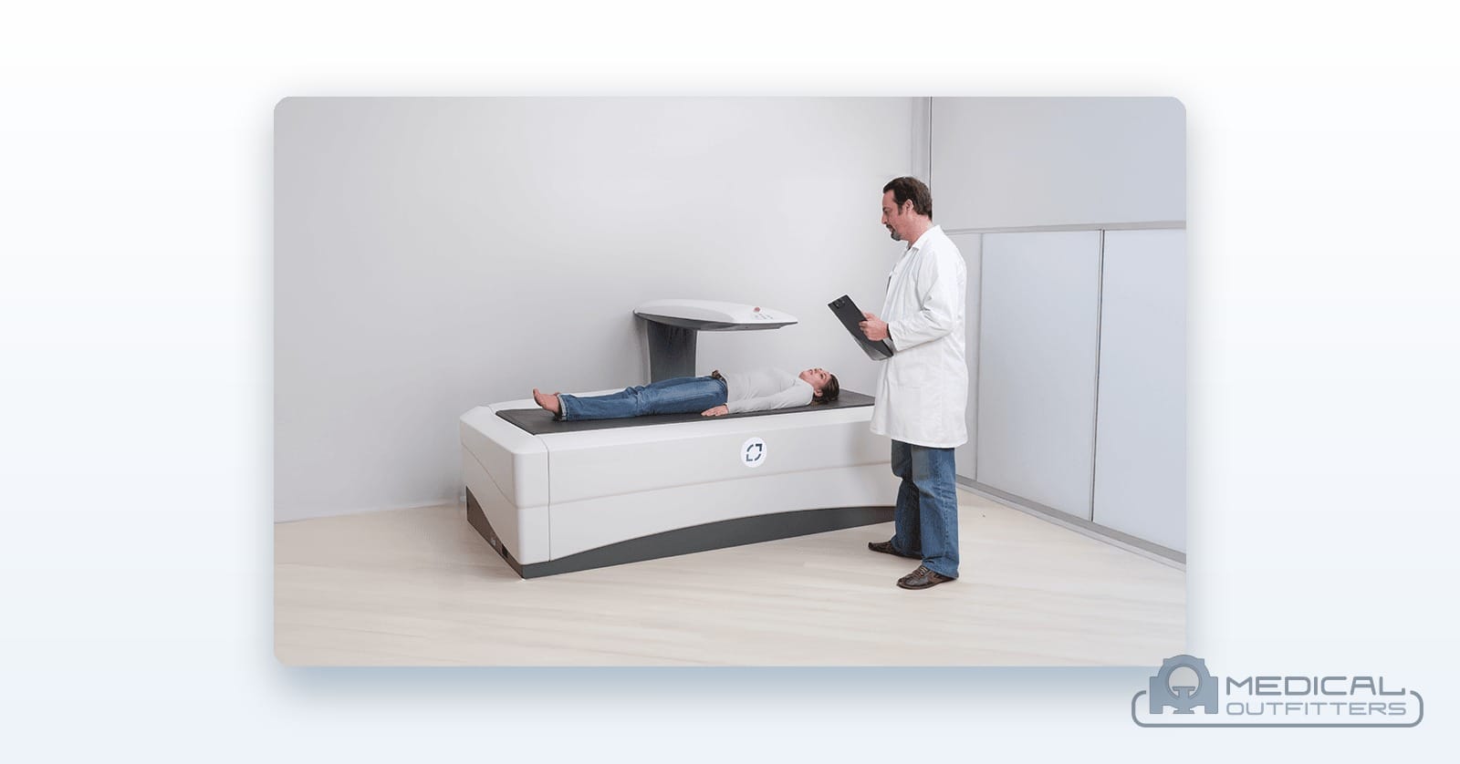
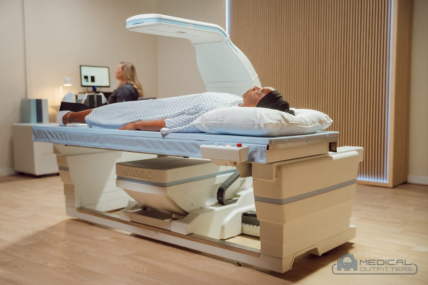
There are two classes of DEXA machine, and the difference matters to both readers of this guide. A central DEXA is a full-size table with a scanning arm that measures the hip, the spine and, on most current systems, the whole body; it is the instrument used for diagnosis and monitoring. A peripheral DEXA measures the forearm, wrist, heel or finger on a compact device. RadiologyInfo notes that peripheral devices are useful for screening but are less useful for monitoring treatment, and that a baseline central DXA is recommended once drug therapy is indicated. When people ask what is a DEXA scan, they almost always mean the central exam, and that is the test the rest of this guide describes.
How Does a DEXA Scan Work?
The trick is in the name: two energies. A DEXA scanner sends X-rays at two different energy levels through the body. Bone and soft tissue absorb the two energies in different proportions, so by comparing how much of each beam reaches the detector the software can subtract the soft tissue and calculate how much mineral is left in the path. The result is reported as areal bone mineral density in grams per square centimeter for each region of interest: the first to fourth lumbar vertebrae (L1 to L4), the total hip and the femoral neck, and, when the hip or spine cannot be used, the one-third radius of the forearm, which are the sites the ISCD’s adult official positions name for diagnosis.
How the beam is shaped is the main engineering difference between machines. A pencil-beam scanner moves one narrow beam and a single detector across the region in a raster pattern; older units took several minutes per site, while current digital pencil beams finish in seconds. A fan-beam scanner spreads the beam over an array of detectors and sweeps the region in one pass, which shortens the scan and makes a lateral image of the spine possible on the same visit. Hologic’s Horizon and GE HealthCare’s Lunar Prodigy and iDXA are fan-beam systems; the DMS Imaging Stratos and Stratos C use what DMS calls a Digital Fast Beam, an enhanced pencil beam, and the Stratos DR uses a 2D fan beam with a 256-element detector, as the DEXA scanner brands comparison sets out.
For the patient, the exam itself is simple. RadiologyInfo describes it as an outpatient test of 10 to 30 minutes with no injection and no anesthesia. For the spine, the patient lies on their back with the legs raised on a padded block so the lower back flattens against the table; for the hip, the foot sits in a brace that turns the hip slightly inward. The arm passes slowly overhead while the patient holds still, sometimes holding a breath for a few seconds. A vertebral fracture assessment, a low-dose lateral image of the spine taken on the same machine, adds only a few minutes, according to RadiologyInfo.
Preparation is light but it matters for accuracy. RadiologyInfo asks patients not to take calcium supplements for at least 24 hours before the exam, to wear loose clothing without metal, and to tell the facility about a recent barium study or a contrast injection for CT or a nuclear medicine scan, because the patient may need to wait 10 to 14 days before a DXA. Each of these leaves material in the scan path that the software would count as bone. Most facilities also hand the patient a short questionnaire about medications, prior fractures and family history, because those answers feed the fracture-risk calculation in the report.
What a DEXA Scan Can Show: T-Score, Z-Score, Fractures and Body Composition
The headline of every bone density report is the T-score. It compares the patient’s bone density with that of a healthy young adult of the same sex at peak bone mass and expresses the gap in standard deviations. The World Health Organization categories, as RadiologyInfo summarizes them, are simple: minus 1 and above is normal, between minus 1 and minus 2.5 is low bone mass, often called osteopenia, and minus 2.5 or lower is osteoporosis. The ISCD’s adult official positions state that the WHO international reference standard is a T-score of minus 2.5 or less at the femoral neck, and that osteoporosis may also be diagnosed in postmenopausal women and men aged 50 and older when the lumbar spine, total hip or femoral neck T-score is minus 2.5 or less, using the lowest of the hip sites.
| Score | Compared with | Result | Source |
|---|---|---|---|
| T-score of minus 1.0 or higher | Healthy young adult, same sex | Normal bone density | WHO categories, via RadiologyInfo |
| T-score between minus 1.0 and minus 2.5 | Healthy young adult, same sex | Low bone mass (osteopenia) | WHO categories, via RadiologyInfo |
| T-score of minus 2.5 or lower | Healthy young adult, same sex | Osteoporosis | ISCD adult official positions |
| Z-score above minus 2.0 | People of the same age and sex | Within the expected range for age | ISCD adult official positions |
| Z-score of minus 2.0 or lower | People of the same age and sex | Below the expected range for age | ISCD adult official positions |
The Z-score answers a different question: how does this patient compare with people of the same age and sex? The ISCD prefers it to the T-score for premenopausal women, men under 50 and children, and describes a Z-score of minus 2.0 or lower as below the expected range for age. A low Z-score is a signal that something other than aging, such as a medication, an endocrine disorder or a nutritional problem, may be taking bone away, and it usually prompts further testing.
Most reports carry more than the two scores. The FRAX tool, developed at the University of Sheffield, combines the femoral neck density with age, sex, weight and clinical risk factors to estimate the 10-year probability of a hip fracture and of a major osteoporotic fracture; the treatment thresholds most US clinicians use for patients with low bone mass, a 3 percent hip or 20 percent major fracture probability, were first set by the National Osteoporosis Foundation, now the Bone Health and Osteoporosis Foundation. A vertebral fracture assessment can find a spine fracture the patient never noticed, which changes the diagnosis on its own. Trabecular bone score, a licensed texture analysis of the spine image, adds information about bone quality on Hologic and GE systems.
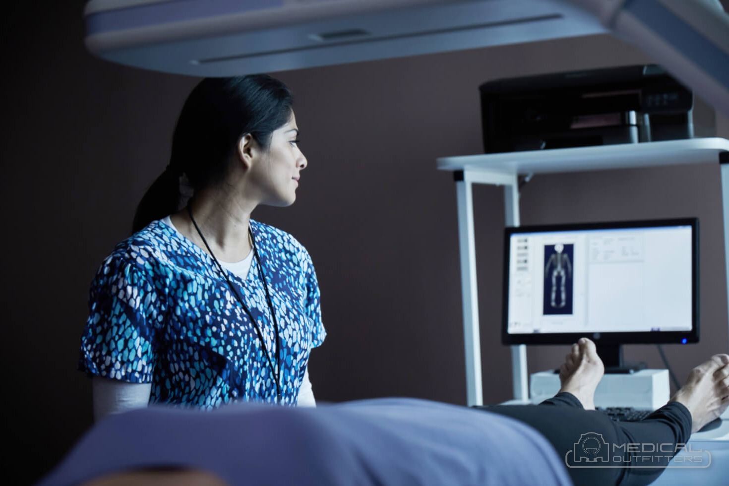
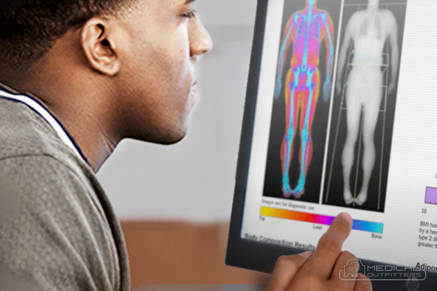
The last thing a DEXA scan can show is body composition. A whole-body scan reports fat mass, lean mass and bone mass for each arm, leg and the trunk, and many systems estimate visceral fat. The ISCD lists monitoring after bariatric surgery or large weight loss, HIV-related fat redistribution and muscle weakness among the clinical indications, and names pregnancy as a contraindication. Outside the clinic, athletes and fitness clients book the same scan to track fat loss and muscle gain, which is why DEXA body composition has become a self-pay service of its own.
How Accurate Is a DEXA Scan? Precision, Least Significant Change and Pitfalls
Two different ideas hide inside the question how accurate is a DEXA scan. Accuracy is how close the measured density is to the true mineral content of the bone; precision is how closely repeated measurements on the same person agree. For a single diagnosis, accuracy matters. For the more common clinical job, telling whether bone density has changed between two scans, precision is what counts, and it is where DEXA earns its place: changes in bone density are small, often a few percent a year, so the test is only useful if its own measurement error is smaller still.
The ISCD sets the bar. Its adult official positions require every facility to run its own precision study, scanning 15 patients three times or 30 patients twice and repositioning the patient between scans, and they state that the manufacturer’s published precision should not be used in its place. The minimum acceptable precision for an individual technologist is 1.9 percent at the lumbar spine, 1.8 percent at the total hip and 2.5 percent at the femoral neck. From those numbers the facility calculates the least significant change (LSC), the smallest difference between two scans that can be called real with 95 percent confidence, which the ISCD glossary defines as the precision error multiplied by 2.77.
In practice that means a follow-up scan has to move by about 5 percent at the spine or hip before the report can call it a true loss or gain. A patient whose spine density falls by 3 percent has not lost bone, as far as the scan can tell; one whose density falls by 6 percent probably has. A careful technologist with a good precision study can report a smaller LSC than the minimum, which is one reason bone density programs invest in positioning training and daily phantom checks.
The pitfalls are well known. RadiologyInfo notes that DXA is less useful in a spine with deformity or prior surgery, that compression fractures and osteoarthritis can affect accuracy, and that CT may be more helpful in those patients: arthritic spurs and hardened joints add mineral to the measured area and push the spine result up, which is why the hip is often the more reliable site in older patients. Calcium tablets, barium and metal in the scan path have the same effect. RadiologyInfo also stresses that follow-up scans should ideally be done at the same facility on the same machine, because results from different equipment cannot be compared directly. The Medical Outfitters brands guide notes that raw BMD values run roughly 10 to 15 percent higher on GE Lunar systems than on Hologic systems, so a patient who changes facilities or a clinic that changes brands needs a new baseline or a formal cross-calibration.
Finally, DEXA measures density, not strength or fate. RadiologyInfo is explicit that the test cannot predict who will fracture; it estimates relative risk. That is the reason modern reports pair the T-score with FRAX and, where licensed, a vertebral fracture assessment and trabecular bone score.
Who Should Have a DEXA Scan, and How Much Radiation Does It Use?
The US Preventive Services Task Force updated its recommendation on January 14, 2025: it recommends osteoporosis screening for women aged 65 and older and for postmenopausal women younger than 65 who are at increased fracture risk, both at grade B, and found the evidence insufficient to recommend for or against routine screening in men. The ISCD’s adult positions go a little further and recommend bone density testing for women aged 65 and older, men aged 70 and older, and younger adults with a risk factor such as low body weight, a prior fracture, a high-risk medication or a disease associated with bone loss. RadiologyInfo’s list of people for whom the test is strongly recommended includes postmenopausal women not taking estrogen, people on long-term corticosteroids, people with type 1 diabetes, hyperthyroidism or hyperparathyroidism, and anyone who has broken a bone after only mild trauma.
Medicare follows the same logic. Medicare Part B covers a bone mass measurement once every 24 months, or more often when medically necessary, for people who meet one of its qualifying conditions, and the patient pays nothing when the provider accepts assignment, as the Medical Outfitters DEXA cost guide summarizes from the Medicare benefit. RadiologyInfo puts routine follow-up at about every two years, with some patients on high-dose steroids needing a repeat at six months.
The DEXA radiation dose is among the lowest in medical imaging. RadiologyInfo’s dose table lists bone densitometry at about 0.001 mSv, comparable to three hours of natural background radiation, against 0.1 mSv for a chest X-ray and 0.28 mSv for a screening digital mammogram. The ISCD’s white paper on precision assessment and radiation safety (Baim and colleagues, Journal of Clinical Densitometry, 2005) puts the effective dose of a spine or hip scan at typically less than 5 microsieverts and a routine spine-and-hip exam at less than 10, close to the roughly 8 microsieverts a person in the United States receives from background radiation every day. That low dose is what makes repeated monitoring practical.
For a facility the low dose has a practical side too. The Medical Outfitters DEXA guide notes that most states register a bone densitometer as an X-ray device and may ask for a shielding evaluation, which the low dose usually satisfies in a standard room, and Hologic’s Horizon specifications state that no external shielding is required. A DEXA room is far simpler to build than a general X-ray room, but the state registration, the physicist’s report and technologist training still belong in the plan.
DEXA Scan Cost in 2026: Cash, Insurance and Medicare
DEXA scan cost is one of the lowest in imaging. CoveredUSA’s national pricing guide of May 15, 2026 puts the national average at $175 without insurance, with most quotes between $80 and $300, a low of about $65 at community screening events and a high of about $600 in a hospital outpatient department. The Medical Outfitters DEXA cost guide, which compiles those surveys with marketplace prices checked on October 12, 2026, puts a typical cash price at $100 to $200 at an independent imaging center or physician office and $200 to $600 at a hospital outpatient department; prepaid marketplace prices at Florida imaging centers ran $91 to $106 on MDsave on that date.
| CPT code | What it bills | 2026 Medicare national amount | Hospital outpatient rate |
|---|---|---|---|
| 77080 | Axial DXA: hips, pelvis and spine, the standard osteoporosis test | $39.41 | $106.81 |
| 77081 | Peripheral DXA: forearm, wrist or heel | $31.73 | $88.91 |
| 77085 | Axial DXA with vertebral fracture assessment | $54.44 | $106.81 |
| 76499 | Unlisted code often used for body composition DEXA | Contractor priced | Usually self-pay |
Source: Medical Outfitters DEXA cost guide, from the CMS Physician Fee Schedule indicators for 2026 at the $33.40 conversion factor and the 2026 outpatient rates in Hologic’s bone densitometry coding guide, drawn from the CMS OPPS addenda.
Insurance follows the Medicare pattern: a bone density test ordered for a clinical reason, such as menopause-related risk, long-term steroid use, a prior fracture or monitoring treatment, is usually covered under the plan’s deductible and copay. A body composition DEXA is the exception. It is self-pay almost everywhere, and the published prices in the Medical Outfitters guide run about $75 to $150 a scan at dedicated body composition clinics, with packages and memberships bringing a repeat scan under $100.
The gap between those prices is the business case on the facility side. Medicare pays a clinic about $39 for an axial study, while cash and commercial patients pay several times that, so a densitometer is a volume purchase. The DEXA cost guide works the arithmetic for a $60,000 refurbished central system: about 1,500 scans to recover the equipment line at $40 a scan, or about 750 at an $80 blended rate, which at 20 scans a week is roughly 17 or 9 months, before technologist time, service and space. For the same view across every modality, see radiology equipment cost in one table.
The Machine Behind the Scan: Hologic Horizon, GE Lunar iDXA and DMS Stratos
Three platforms account for almost every central DEXA a US clinic will consider. Hologic’s Horizon family is the current generation from the maker the Medical Outfitters brands guide calls the US market leader: the A, W and C tables with high-definition vertebral fracture assessment (IVA HD) and the lighter Wi and Ci configurations with standard IVA, all fan-beam, with a 500 pound (227 kg) patient weight limit and no external shielding required, according to Hologic’s Horizon technical specifications. The previous Discovery A, W and C series is the backbone of the refurbished market.
GE HealthCare’s Lunar line runs from the legacy DPX pencil-beam units through the Prodigy, the most common refurbished central DEXA in circulation per the Medical Outfitters guide, to the iDXA, the premium narrow-fan-beam platform with a direct-digital detector and advanced body composition. GE’s iDXA specification sheet lists a 450 pound (204 kg) maximum patient weight. The third is DMS Imaging’s Stratos line, which Medical Outfitters sells new as the authorized dealer: the Stratos with its Digital Fast Beam, the fan-beam Stratos DR at about 15 seconds per site with body composition and 3D-DXA femur modeling, and the compact Stratos C for a small room. The Stratos series carries the CE mark under the EU Medical Device Regulation; FDA clearance is pending, per the Medical Outfitters guide, so the regulatory status for a US site is confirmed with every quote.
| DEXA system class | Refurbished, all-in | New, all-in |
|---|---|---|
| Peripheral DEXA (forearm or heel) | $18,000 to $40,000 | $40,000 to $70,000 |
| Central, full-body DEXA | $35,000 to $85,000 | $85,000 to $165,000 |
| GE Lunar iDXA | $72,000 to $85,000 | Estimated through the OEM: $125,000 to $165,000 |
| Hologic Horizon W | $70,000 to $82,000 | Estimated through the OEM: $110,000 to $145,000 |
| DMS Stratos DR | Sold new only | $125,000 to $165,000 |
Source: Medical Outfitters DEXA and bone densitometer cost guide, all-in ranges delivered, installed and warrantied. OEM new figures for GE and Hologic are comparison estimates and not Medical Outfitters offers.
The choice between them comes down to four questions. Does the program need body composition, or only bone density? Which reference database do the reading physicians know, given that a clinic changing brands has to re-baseline its patients? What is the patient population, including the heaviest patient the table must hold? And who will service the system locally? A refurbished Hologic Discovery or GE Prodigy suits a clinic adding bone density on a budget; a Horizon W, an iDXA or a Stratos DR suits a practice that sells body composition as a service. Refurbished systems in the MO Select program are inspected, calibrated against a phantom and installed with a written warranty, as the refurbished imaging equipment page describes, and the full model list is in the bone densitometry category.
Adding Bone Density Testing With Medical Outfitters
A DEXA scan is a short, low-dose test with a long reach: it diagnoses osteoporosis, tracks treatment, finds silent spine fractures and, on the same table, measures body composition. For a clinic, the densitometer is one of the most affordable imaging systems to add and one of the fastest to pay back when the referral base is there. Start with the DEXA and bone densitometer cost guide for the price bands and the payback calculator, compare platforms in DEXA scanner brands and what to check when buying used, and look at the new DMS Stratos DR and the compact Stratos C. For installation and support, see installation and rigging and service and repairs. When you know your expected volume and payer mix, send it to us and we will price a specific system.
- RadiologyInfo (ACR and RSNA), Bone Densitometry (DEXA, DXA): sites scanned, exam length, preparation, T-score and Z-score, limitations, follow-up: https://www.radiologyinfo.org/en/info/dexa
- RadiologyInfo, Radiation Dose in X-Ray and CT Exams: 0.001 mSv for bone densitometry and the comparison doses: https://www.radiologyinfo.org/en/info/safety-xray
- International Society for Clinical Densitometry, Adult Official Positions: diagnostic sites, Z-score wording, precision minimums and LSC, testing indications, body composition: https://iscd.org/learn/official-positions/adult-positions/
- Baim S. et al., Precision Assessment and Radiation Safety for DXA, ISCD white paper, Journal of Clinical Densitometry 2005: effective doses and background comparison: https://iscd.org/wp-content/uploads/2026/04/Radiation-Safety-White-Paper.pdf
- US Preventive Services Task Force, Screening for Osteoporosis to Prevent Fractures, final recommendation January 14, 2025: https://www.uspreventiveservicestaskforce.org/uspstf/recommendation/osteoporosis-screening
- Bone Health and Osteoporosis Foundation, Osteoporosis Fast Facts: prevalence and lifetime fracture risk: https://bonehealthandosteoporosis.org/wp-content/uploads/Osteoporosis-Fast-Facts-2.pdf
- Hologic, Horizon DXA System technical specifications (2024 edition) and skeletal health product pages: models, patient weight limit, shielding: https://www.hologic.com/skeletal-health
- Medical Outfitters, DEXA and Bone Densitometer Cost and Price Guide (2026): scan prices, CPT rates, machine bands, payback: https://medicaloutfitters.com/dexa-bone-densitometer-cost-guide/
- DEXA and bone densitometer cost guide (2026)
- DEXA scan machines and bone densitometers for sale
- DEXA scanner brands compared and buying used
- DMS Stratos DR bone densitometer
- DMS Stratos bone densitometer
- DMS Stratos C compact densitometer
- Refurbished medical imaging equipment: the MO Select standard
- Radiology equipment cost in 2026, every modality
- X-ray room size and requirements
- Installation, rigging and de-installation
- Imaging equipment service and repairs
- Price a DEXA system for your clinic
DEXA Scans: Common Questions
What is a DEXA scan?
A DEXA scan, short for dual-energy X-ray absorptiometry and also written DXA, is a low-dose X-ray test that measures bone mineral density, usually at the lower spine and hips. RadiologyInfo calls it the standard method for diagnosing osteoporosis. The exam takes 10 to 30 minutes, uses about 0.001 mSv of radiation, roughly three hours of natural background, and reports the result as a T-score and a Z-score.
How does a DEXA scan work?
The scanner sends X-rays at two energy levels through the body. Bone and soft tissue absorb the two energies in different proportions, so the software subtracts the soft tissue and calculates the mineral left in the path, in grams per square centimeter. The ISCD names the lumbar spine (L1 to L4), the total hip and the femoral neck as the diagnostic sites, with the one-third radius of the forearm when the hip or spine cannot be used.
What can a DEXA scan show?
Bone mineral density as a T-score against a healthy young adult, where minus 2.5 or lower means osteoporosis, and a Z-score against people of the same age and sex. Most reports add a FRAX 10-year fracture probability. On current systems the same table can take a vertebral fracture assessment, which finds spine fractures the patient never noticed, and a whole-body scan that reports fat, lean and bone mass by region.
How accurate is a DEXA scan?
Precise enough to track change when the same machine is used. The ISCD requires each facility to measure its own precision, with minimums of 1.9 percent at the spine, 1.8 percent at the total hip and 2.5 percent at the femoral neck, which gives a least significant change of 5.3, 5.0 and 6.9 percent. Spinal arthritis, compression fractures, calcium tablets and metal can raise the spine result, and scans from different brands cannot be compared directly.
Is a DEXA scan safe?
Yes, for nearly everyone. RadiologyInfo lists the dose at about 0.001 mSv, against 0.1 mSv for a chest X-ray, and the ISCD's radiation safety white paper puts a routine spine and hip exam under 10 microsieverts, similar to one day of natural background radiation in the United States. No radiation stays in the body afterward. Pregnancy should be reported first, and the ISCD lists pregnancy as a contraindication to a body composition DEXA.
Who should have a DEXA scan?
The US Preventive Services Task Force recommended in January 2025 that women aged 65 and older, and postmenopausal women under 65 at increased fracture risk, be screened for osteoporosis. The ISCD also recommends testing men aged 70 and older and younger adults with risk factors such as low body weight, a prior fragility fracture, long-term steroids or a disease linked to bone loss. Medicare Part B covers the test once every 24 months for qualifying patients.
How much does a DEXA scan cost?
About $100 to $200 cash at an independent imaging center and $200 to $600 at a hospital outpatient department, per the Medical Outfitters DEXA cost guide; CoveredUSA's May 2026 guide puts the national average at $175. Medicare Part B covers a qualifying patient at $0 every 24 months, and the 2026 Medicare national amount for CPT 77080 is $39.41. A body composition DEXA is self-pay, about $75 to $150 a scan at dedicated clinics.
How much does a DEXA machine cost?
The Medical Outfitters DEXA cost guide puts a central, full-body densitometer at $35,000 to $85,000 refurbished and $85,000 to $165,000 new, and a peripheral forearm or heel unit at $18,000 to $40,000 refurbished and $40,000 to $70,000 new, delivered, installed and warrantied. Hologic Horizon and GE Lunar systems come refurbished on request through MO Select; the DMS Stratos line is sold new, with its US regulatory status confirmed on every quote.
Related Articles