So what is fluoroscopy, and how is it different from the X-ray that takes a single picture? A standard radiograph is one frozen exposure; fluoroscopy is the live, moving version of the same physics, a stream of X-ray images shown in real time so the operator can watch a process happen and act on it. It is how a surgeon steers a wire into an artery, how a radiologist follows barium down the throat, and how an orthopedist confirms a fracture is set before the patient leaves the table. This guide explains what fluoroscopy is, how the live image is made, the difference between pulsed and continuous beams and between the image intensifier and the flat panel detector, the three places fluoroscopy is done, the common exams, how the radiation dose is kept in check, and what the equipment costs.
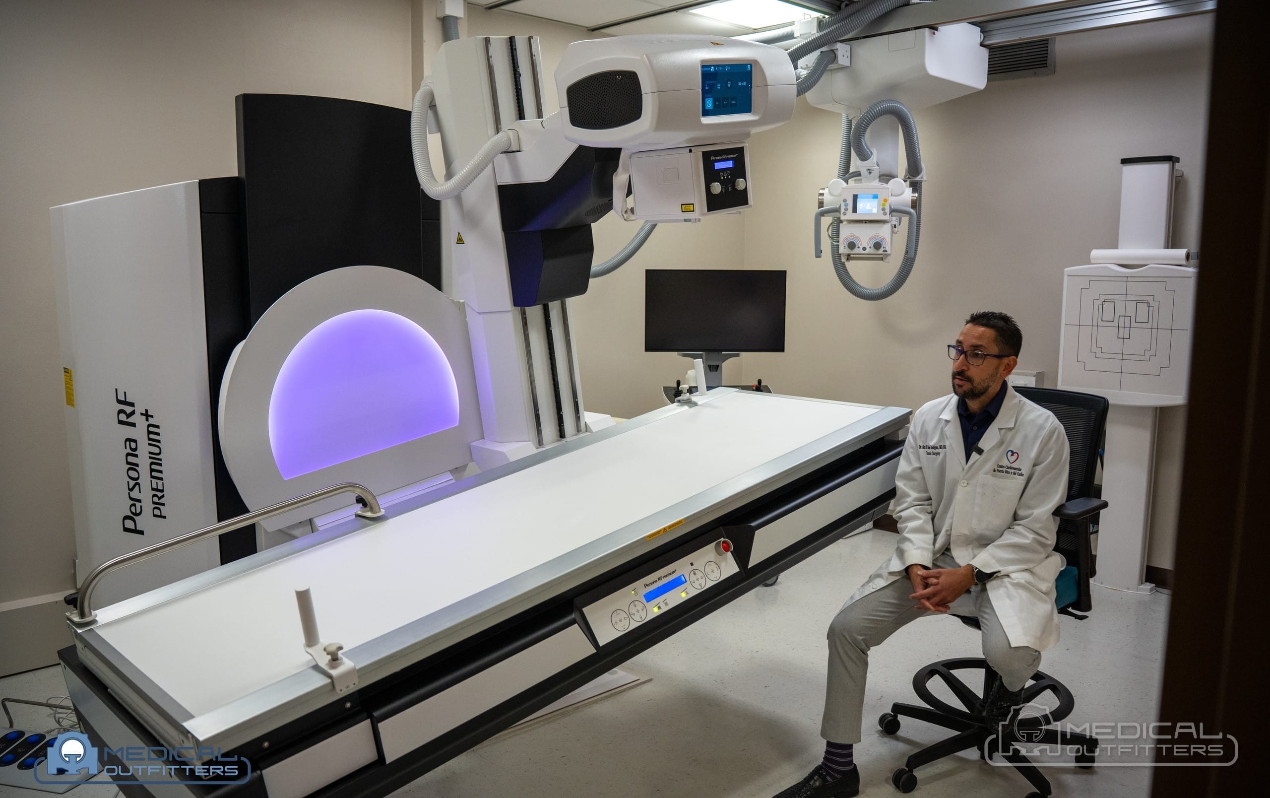
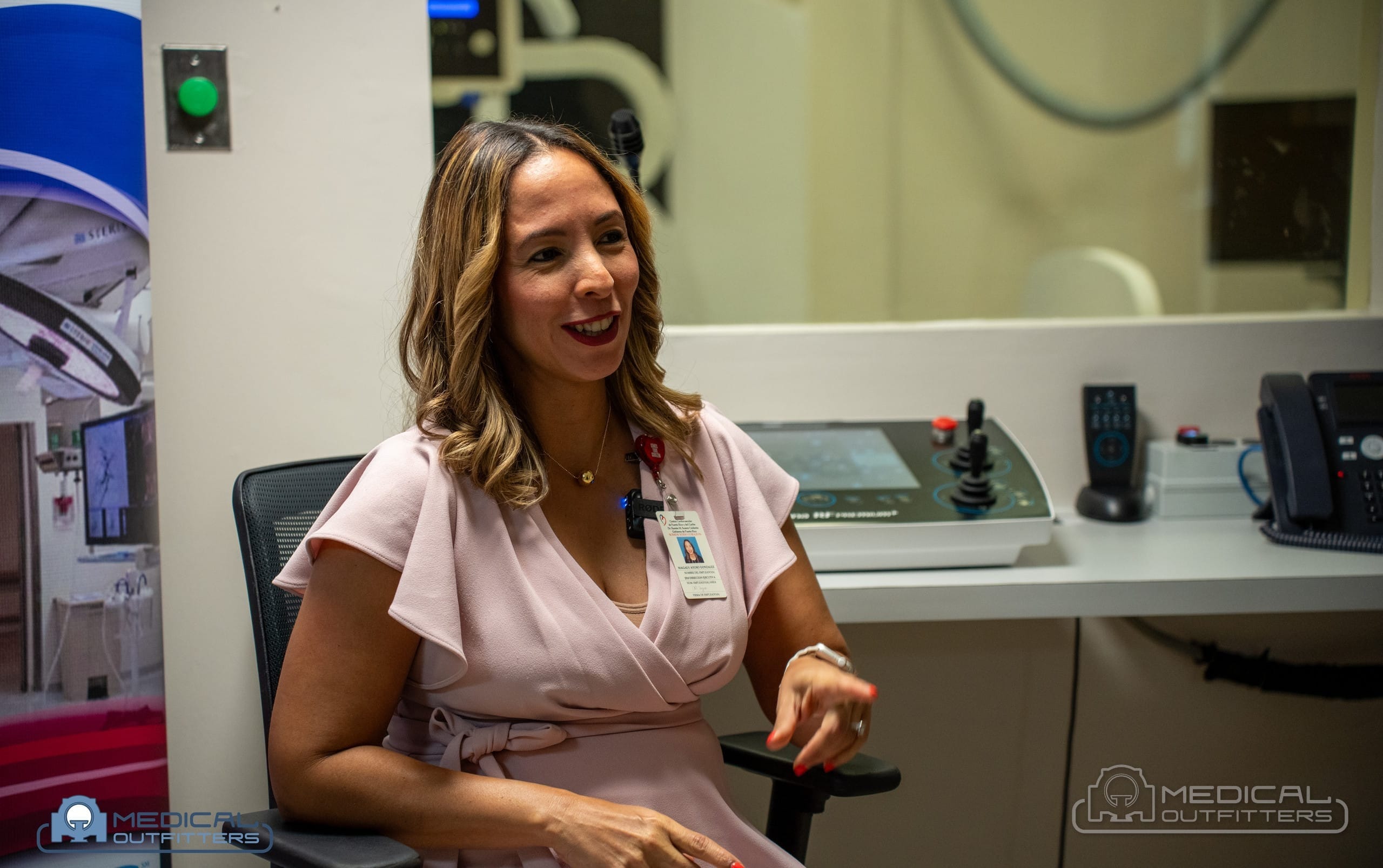
Medical Outfitters sells and installs the systems that do fluoroscopy, from mobile C-arms to fixed radiography and fluoroscopy rooms such as the Fujifilm Persona RF the company installed for Centro Cardiovascular de Puerto Rico y del Caribe, and services them afterward. This article is written for the facility owner, radiology manager or physician deciding what kind of fluoroscopy capability a practice needs and what it will cost, not for the patient on the table, though it answers the patient’s questions too.
- What it is: a continuous, moving X-ray image shown live, used to guide procedures and watch contrast move, not to take one still picture.
- How it is made: pulsed or continuous X-rays strike a flat panel detector or an image intensifier, which turns them into the live video on the monitor.
- Where it is done: fixed radiography and fluoroscopy (R/F) rooms, mobile C-arms in the operating room and clinic, and fixed interventional and cath lab suites.
- What it is used for: barium and swallow studies, catheter and line placement, pain and joint injections, urology studies, and orthopedic and vascular work.
- Dose and cost: the dose is higher than a plain X-ray and managed by pulsing and good technique; the machines run from tens of thousands of dollars for a used C-arm to a per-project price for a fixed room.
What Is Fluoroscopy? From Still X-Ray to Moving Image
The FDA defines fluoroscopy as a type of medical imaging that “shows a continuous X-ray image on a monitor, much like an X-ray movie.” That one sentence holds the whole difference from plain radiography. A chest or bone X-ray is a single exposure: the tube fires once, the detector records one frame, and the study is a still picture. In fluoroscopy the tube keeps producing X-rays, the detector keeps reading them out, and the result is live video of what the beam passes through, updated many times a second while the clinician watches and moves instruments or the patient.
That live feedback is the point. A still X-ray shows where something is; fluoroscopy shows what is happening and lets the operator respond to it in the moment. A guidewire is advanced a centimeter and the screen confirms it went the right way; contrast is injected and the screen shows it fill a vessel, a joint or the bowel; a fracture is manipulated and the screen shows the fragments line up. Because the beam is on while all of this happens, fluoroscopy delivers more radiation than a single radiograph, which is why dose management, covered below, is built into both the equipment and the regulations. The receptor technology behind both still and moving X-ray is the same family of flat panels described in our guide to CR vs DR X-ray.
How the Live Image Is Made: Pulsed vs Continuous
A fluoroscopy system is an X-ray tube on one side of the patient and a detector on the other, with a control that the operator holds down to make the beam. Under 21 CFR 1020.32, the system will only expose while the operator keeps continuous pressure on that control, so the beam is on only when it is being watched, and the time counts from the first instant of pressure to the last.
Early fluoroscopy ran the beam continuously, like a film camera with the shutter always open. Modern systems pulse instead: the tube fires brief bursts, often at 3, 7.5, 15 or 30 pulses a second, and the screen holds the last frame between pulses so the image looks continuous to the eye. Pulsing is the single biggest dose lever on the machine, because a procedure that can be guided at 7.5 pulses a second uses far less radiation than one run continuously, and the operator or a protocol chooses the rate for the task. The FDA sets the ceiling: under the air kerma rate limits in 21 CFR 1020.32, normal fluoroscopy may not exceed roughly 88 milligray per minute at the reference point, about 10 roentgens per minute, and a separate high-level control mode that allows more is permitted only under continuous manual activation with an audible signal sounding the whole time.
Between the pulses, two features cut dose further without the operator losing the picture. Last image hold freezes the final frame on the screen so the team can study anatomy with the beam off, and the dose readout on the console, the reference air kerma and the kerma area product, tells the operator how much radiation the study has used so far. Those two numbers are the ones the FDA asks facilities to record for every fluoroscopic procedure.
Image Intensifier vs Flat Panel Detector
What turns the X-rays into the picture on the screen is the detector, and there are two generations of it. The older one is the image intensifier, a large vacuum tube that converts X-rays to light, amplifies that light electronically and feeds a video camera. It served fluoroscopy for decades and still works well, especially in its magnified modes, but it is bulky, and the RSNA and AAPM physics tutorial on flat-panel fluoroscopy notes that image intensifiers suffer from glare, vignetting that darkens the edges, and geometric distortions that bend a straight line near the rim of the field.
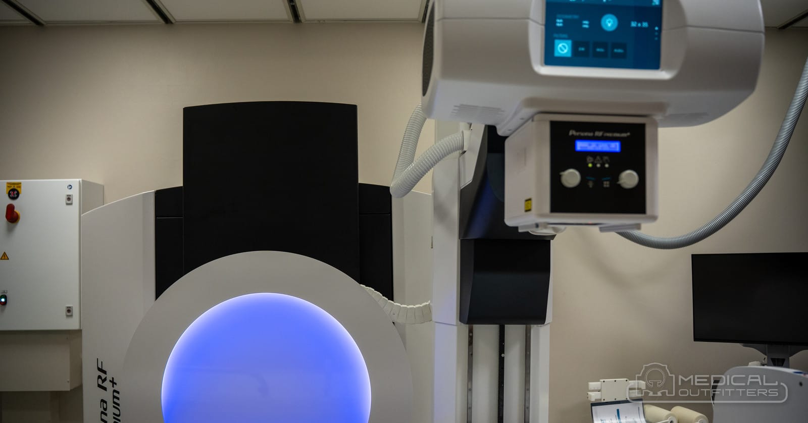
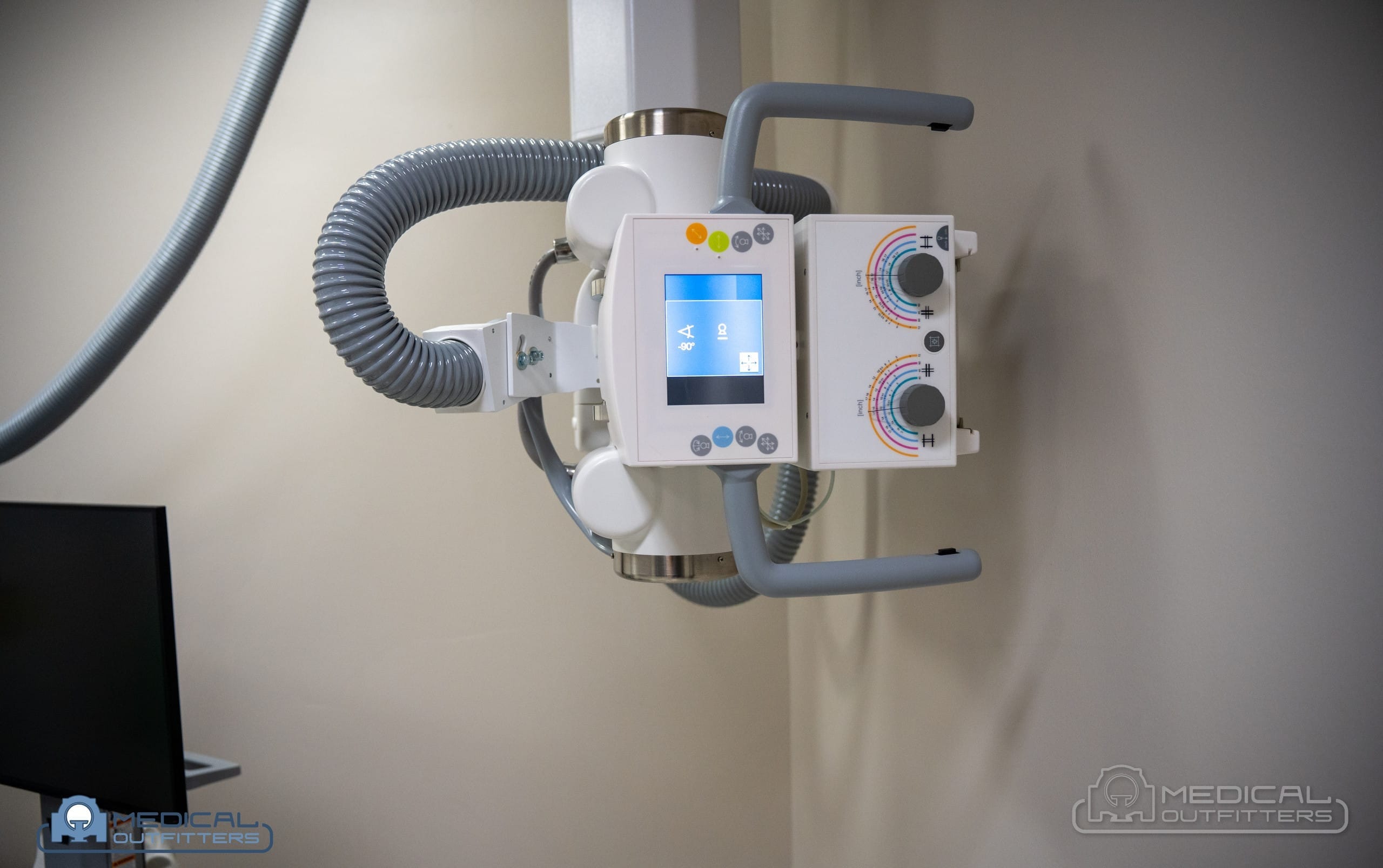
The newer one is the dynamic flat panel detector, the same flat-panel technology used in digital radiography but read out fast enough for video. The flat panel is thin, so it fits a mobile C-arm or a compact room, and the same tutorial notes that it has none of the image intensifier’s geometric distortion and a wider dynamic range, so the picture is even across the whole field and holds detail in both bright and dark areas. A straight catheter looks straight at the edge of the screen, not curved. Most systems sold today, from mobile C-arms to fixed rooms, use a flat panel; the Fujifilm Persona RF room, for example, is built around a 17 by 17 inch, 140 micron indium gallium zinc oxide dynamic flat panel, per Fujifilm’s product specification.
Where Fluoroscopy Is Done: R/F Rooms, C-Arms and Interventional Suites
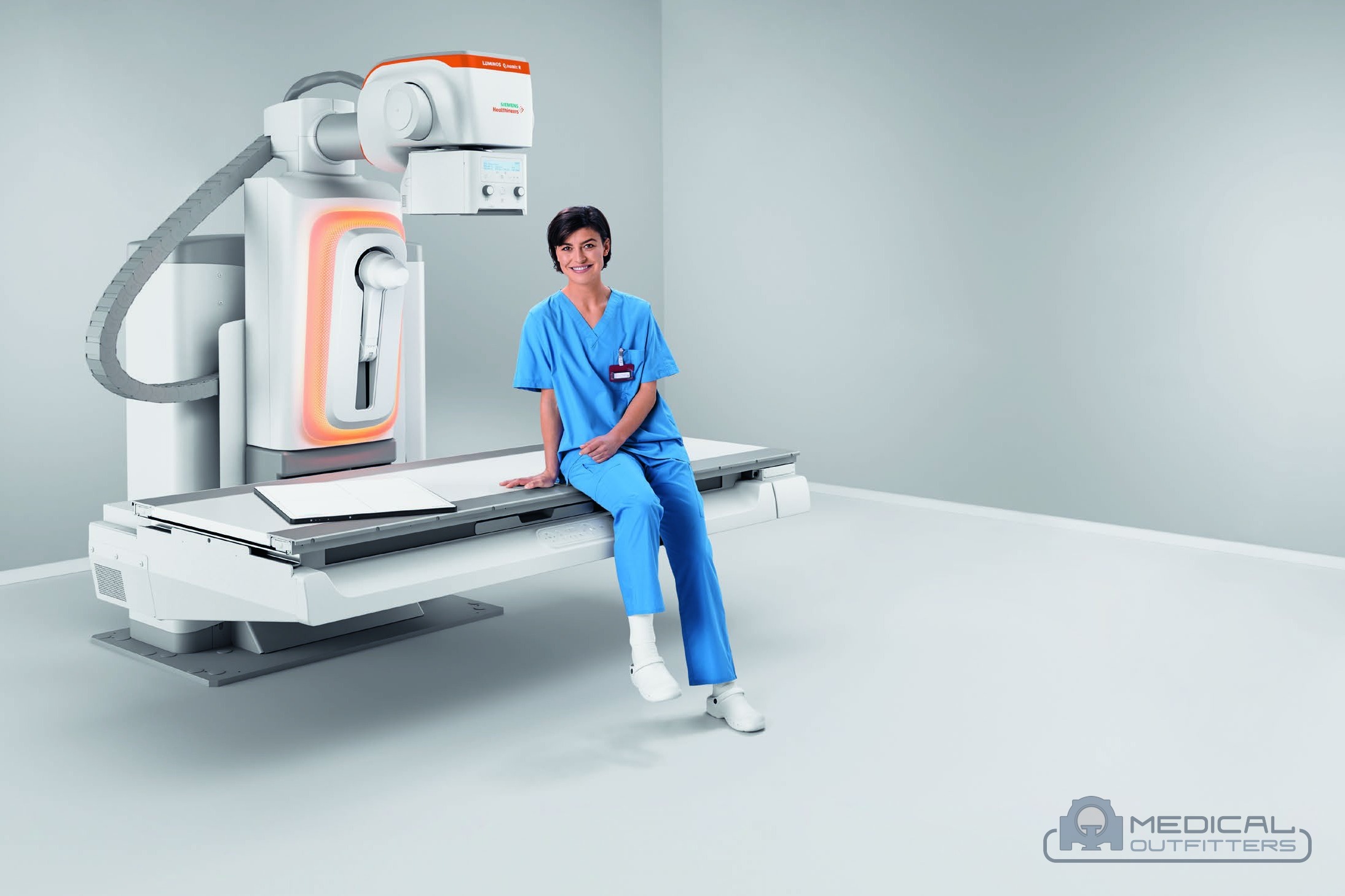
Fluoroscopy is the method; the fluoroscopy machine takes three main shapes, and which one a facility needs depends on the work. The first is the fixed radiography and fluoroscopy room, the R/F room, a shielded radiology room with an over- or under-table tube, a tilting table and a flat panel, that does both ordinary X-rays and fluoroscopic studies from one console. This is the home of barium and swallow studies and general fluoroscopy; the Fujifilm Persona RF and the Siemens LUMINOS line are examples, the Persona RF switching on demand between radiographic, fluoroscopy and tabletop exams with a table that tilts a full plus and minus 90 degrees and up to a 71 inch source-to-image distance, per Fujifilm.
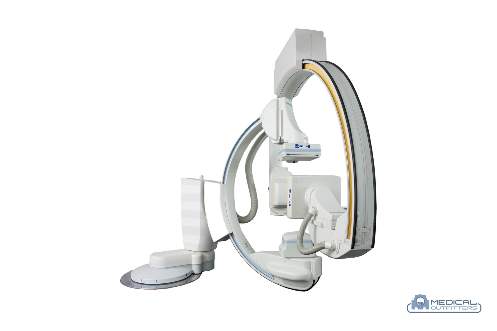
The second is the mobile C-arm, a wheeled unit with the tube and detector on a C-shaped arm that rolls into an operating room, a pain clinic or an orthopedic suite and images from almost any angle. It is the fluoroscopy most clinicians meet, and it ranges from a small mini C-arm for hands and feet to a full-size surgical unit and a 3D vascular system; the full comparison is in our guide to the difference between a C-arm and fluoroscopy and in the C-arm machine cost guide. The third is the fixed interventional or cardiac catheterization suite, a built-in, often floor- or ceiling-mounted system with a powerful tube, advanced software and sometimes two planes at once, used for angiography, cardiac and neurovascular work; that room is covered in our guide to what a cath lab is. All three do fluoroscopy; they differ in power, mobility, image size and price.
What Fluoroscopy Is Used For: The Common Procedures
The FDA lists fluoroscopy’s uses as a wide range of diagnostic and treatment procedures, among them barium X-rays, catheter insertion, stent placement, angiograms and orthopedic surgery. In practice they fall into a few groups. Contrast studies of the gut use swallowed or instilled barium watched live: the barium swallow and upper GI series for the esophagus and stomach, the modified barium swallow run with a speech pathologist to study swallowing, and lower GI and small-bowel studies. Interventional and vascular work uses fluoroscopy to steer catheters, wires, stents and filters through vessels, to place central lines and feeding tubes, and to perform angiography and biliary procedures such as ERCP.
Pain management and orthopedics use it for guided spinal injections, facet and epidural injections, joint injections and arthrograms, and to confirm the reduction of a fracture or the placement of hardware in surgery. Urology and gynecology use it for voiding studies of the bladder and for tubal studies. What ties them together is that the clinician needs to see motion or guide an instrument in real time, which a single still X-ray cannot provide. For a facility, the mix of these exams decides the equipment: a GI and general practice wants a fixed R/F room, a surgery center or pain clinic wants a mobile C-arm, and a cardiology or vascular program wants a fixed interventional suite.
Radiation Dose and How It Is Managed
Because the beam is on while the procedure happens, fluoroscopy uses more radiation than a plain X-ray, and the FDA is direct about it: fluoroscopy “can result in relatively high radiation doses, especially for complex interventional procedures,” with the main risks being skin and tissue injuries that can appear after a long procedure and a small long-term cancer risk. The doses vary enormously with the exam. A diagnostic study is modest: a modified barium swallow averaged 0.32 millisievert of effective dose across 200 adults in a 2021 Dysphagia study, and the Health Physics Society puts a barium swallow near 1 millisievert and an upper GI series near 6, against roughly 0.1 millisievert for a chest X-ray. A long interventional case can deliver far more, which is why those cases are the ones the FDA singles out.
The dose is managed by the machine and by the operator. On the machine, the pulsed beam, last image hold, dose-rate controls and spectral filters hold the radiation down, and the console shows the running reference air kerma and kerma area product so the team knows where it stands. By the operator, the rule is the one the FDA states, the “lowest acceptable exposure for the shortest time necessary,” achieved by keeping the beam off except when it is being used, keeping the detector close to the patient, collimating tightly, using the lowest pulse rate that still guides the work, and watching the dose readout. For the staff, lead aprons, thyroid shields, ceiling-mounted shields and distance from the beam are the protection, and the equipment Medical Outfitters installs is delivered with these dose features configured and, through our X-ray service team, kept calibrated so the output on the display matches the output at the table.
What Fluoroscopy Equipment Costs
A fluoroscopy machine is priced by its class, not by the word fluoroscopy, and the clearest way to read the market is through the equipment that does it. Mobile C-arms are the priceable core: the Medical Outfitters C-arm machine cost guide gives all-in bands, delivered, installed and warrantied, of $45,000 to $85,000 refurbished and an estimated $140,000 to $185,000 new for a mini or extremity C-arm, $55,000 to $135,000 refurbished and $165,000 to $295,000 new for a full-size mobile C-arm, and $125,000 to $240,000 refurbished and an estimated $275,000 and up new for a 3D or premium vascular C-arm, with a full-size unit renting at roughly $2,500 to $7,000 a month. A fixed R/F room and a fixed interventional suite are quoted per project, because the price depends on the room build, the shielding and the power as much as on the system.
| Fluoroscopy system class | Where it is used | Refurbished, all-in | New, all-in |
|---|---|---|---|
| Mini / extremity C-arm | Hands, wrists, feet, ankles | $45,000 to $85,000 | Not sold new by Medical Outfitters; estimated at $140,000 to $185,000, comparison only |
| Full-size mobile C-arm | Operating room, pain, ortho | $55,000 to $135,000 | $165,000 to $295,000 |
| 3D or vascular C-arm | Vascular, spine, hybrid use | $125,000 to $240,000 | Not sold new by Medical Outfitters; estimated at $275,000 and up, comparison only |
| Fixed R/F room | GI, swallow, general fluoro | Quoted per project, plus room build and shielding | |
| Interventional / cath lab suite | Cardiac, vascular, neuro | Quoted per project; see the cath lab guide | |
The figures are Medical Outfitters planning bands from the C-arm machine cost guide, not quotes, and a written quotation for a specific unit may differ. What a buyer actually weighs is the fit: a clinic doing extremity work does not need a vascular system, and a GI practice is better served by a fixed R/F room than by a C-arm. The X-ray machine cost guide and the radiology equipment cost overview place these numbers next to the other modalities a facility is budgeting at the same time.
Fluoroscopy Equipment With Medical Outfitters
Medical Outfitters supplies and installs the full range of fluoroscopy equipment: mobile C-arms new and refurbished, listed in the C-arm category and priced in the C-arm cost guide, with C-arm rental for short-term needs; fixed radiography and fluoroscopy rooms such as the digital fluoroscopy rooms built around the Fujifilm Persona RF, like the one installed for Centro Cardiovascular de Puerto Rico; and the interventional and cardiac rooms described in our cath lab guide. Every system is delivered with its dose features configured, and our X-ray service team keeps the output calibrated and the system within spec afterward. For the detector side of digital imaging see CR vs DR X-ray, and to match a fluoroscopy system to your exam mix and room, contact Medical Outfitters.
- US FDA, Fluoroscopy: definition, uses, dose and risk, and dose management. fda.gov
- US FDA, 21 CFR 1020.32, Fluoroscopic equipment: continuous activation and air kerma rate limits. ecfr.gov
- RSNA and AAPM, Physics of Flat-Panel Fluoroscopy Systems, RadioGraphics 2011: image intensifier vs flat panel. pubs.rsna.org
- Tipnis et al., Radiation Effective Doses to Adults Undergoing Modified Barium Swallow Studies, Dysphagia 2021. pubmed.ncbi.nlm.nih.gov
- Health Physics Society, Radiation Exposure From Medical Exams and Procedures: barium swallow and upper GI doses. hps.org
- Fujifilm, Persona RF PREMIUM radiography and fluoroscopy room specifications. fujifilm.com
- Medical Outfitters, C-arm machine cost and buying guide: fluoroscopy equipment bands. medicaloutfitters.com
- C-arm machine cost and buying guide (2026)
- C-arm machines: new and refurbished
- C-arm rental: cost, terms and how it works
- Digital fluoroscopy rooms
- X-ray machine cost and price guide (2026)
- X-ray machine service, repair and calibration
- C-arm vs fluoroscopy: what is the difference
- What is a cath lab? Equipment, room and cost
- CR vs DR X-ray: how the detectors differ
- A Fujifilm Persona RF fluoroscopy room in Puerto Rico
- Radiology equipment cost in 2026, every modality
- X-ray machines for sale: new and refurbished
- X-ray machines: rooms, portables and R/F systems
- Match a fluoroscopy system to your exam mix
Fluoroscopy: Common Questions
What is fluoroscopy?
Fluoroscopy is a form of medical imaging that produces a continuous, moving X-ray image on a monitor, which the FDA describes as much like an X-ray movie. Instead of a single still radiograph, the X-ray beam runs, usually in short pulses, while the clinician watches live and guides a catheter, follows swallowed contrast or positions a bone. It is the real-time version of an ordinary X-ray, used to watch a process and act on it as it happens.
What is the difference between fluoroscopy and an X-ray?
A plain X-ray is a single exposure that records one still picture; fluoroscopy keeps the beam producing images, so the result is live, moving video. The physics is the same, but the still shows where something is while fluoroscopy shows what is happening and lets the operator respond. Because the beam is on longer, fluoroscopy delivers more radiation than one radiograph, so it is reserved for tasks that need real-time guidance rather than a single picture.
What is a fluoroscopy machine?
A fluoroscopy machine is an X-ray tube and a detector, with the patient between them, and a control the operator holds to make the beam. It takes three main forms: a fixed radiography and fluoroscopy room with a tilting table, a mobile C-arm that rolls into an operating room or clinic, and a fixed interventional suite for cardiac and vascular work. All three produce the same live image; they differ in power, size, mobility and price.
What is pulsed fluoroscopy?
Pulsed fluoroscopy fires the X-ray tube in brief bursts, often 3, 7.5, 15 or 30 times a second, rather than running the beam continuously, and the screen holds the last frame between pulses so the image still looks smooth. Pulsing is the biggest dose-saving feature on the machine, because a lower pulse rate uses less radiation for the same guidance. The operator or a stored protocol picks the rate to match the task at hand.
What is the difference between an image intensifier and a flat panel detector?
Both turn X-rays into the picture, by different means. An image intensifier is a large vacuum tube that converts X-rays to light and amplifies it for a camera; it works well but is bulky and, per the RSNA and AAPM physics tutorial, shows glare, edge darkening and geometric distortion. A dynamic flat panel detector is thin, free of that geometric distortion and has a wider dynamic range, which is why nearly all systems sold today use one.
What is a fluoroscopy procedure used for?
Fluoroscopy guides any task that needs real-time imaging. The FDA lists barium X-rays, catheter insertion, stent placement, angiograms and orthopedic surgery among its uses. In practice that means contrast studies of the gut such as the barium swallow and upper GI series, catheter and line placement and angiography, pain and joint injections and arthrograms, orthopedic fracture reduction, and urology studies of the bladder. The common thread is watching motion or steering an instrument as it happens.
How much radiation does fluoroscopy use?
More than a plain X-ray, because the beam is on during the procedure, and it varies widely by exam. A modified barium swallow averaged 0.32 millisievert in a 2021 Dysphagia study, the Health Physics Society puts a barium swallow near 1 millisievert and an upper GI near 6, and a chest X-ray is about 0.1. A long interventional case can deliver much more, which is why the FDA calls out complex procedures and stresses the lowest dose for the shortest time.
How much does a fluoroscopy machine cost?
By class, per the Medical Outfitters C-arm machine cost guide, all-in and installed: a mini C-arm runs $45,000 to $85,000 refurbished and an estimated $140,000 to $185,000 new, a full-size mobile C-arm $55,000 to $135,000 refurbished and $165,000 to $295,000 new, and a 3D or vascular C-arm from $125,000 refurbished into the mid six figures new. A fixed R/F room or interventional suite is quoted per project, because the room build and shielding drive the price.