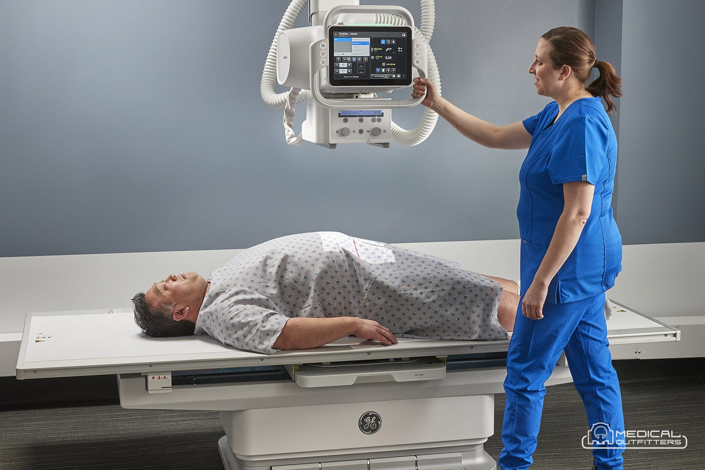
A digital X ray machine is the standard radiography purchase in 2026, whether it is a fixed room with a ceiling-mounted tube, a motorized cart that goes to the ward, or a handheld unit carried to a bedside. The name covers a family of products that share four parts: a generator that makes the high voltage, an X-ray tube on a stand, a flat-panel detector that turns the X-rays into an image, and a workstation that processes the image and sends it to PACS. The buying decisions sit inside those parts: a cesium iodide panel or a gadolinium one, a wireless detector or a fixed one, a 32 kW generator or an 80 kW one, a floor-mounted column or an overhead crane. This guide explains what a digital X-ray machine is, how the detector works, what the generator and the stand decide, the types of system from room to handheld, the software, the dose, and what each class costs, with every figure sourced.
Medical Outfitters sells new Fujifilm digital radiography rooms, portables and wireless FDR D-EVO III detectors as an authorized dealer, sources refurbished GE, Siemens and other DR rooms through MO Select, retrofits flat panels into working film and CR rooms, and installs and services all of it with its own crews across the United States, Puerto Rico and the Caribbean. The guide is written for the administrator, practice owner or radiology manager who has to specify and budget the system, and for the clinician who wants to know what the terms on the quote mean.
- The definition: a digital radiography (DR) system captures the image on a flat-panel detector and shows it in seconds; computed radiography (CR) exposes a cassette that is scanned later, and Medicare pays CR 10 percent less on the technical component.
- The detector: cesium iodide panels convert more of the beam into image than gadolinium oxysulfide panels, about 1.5 times the detective quantum efficiency in a published bench comparison, which buys dose or image quality.
- The generator and stand: a 32 kW generator serves a clinic room and 65 to 82 kW a hospital room; the stand (column, straight arm, U-arm or ceiling crane) sets the room tier and most of the price gap.
- The types: fixed DR rooms, full-size mobile carts, compact carts, handheld units, DR retrofits of existing rooms, and radiography and fluoroscopy rooms, each in its own price band.
- The cost: $12,000 to $65,000 for a portable or handheld unit, $40,000 to $185,000 for a mobile cart and $60,000 to $275,000 for a fixed room, all-in, per the Medical Outfitters X-ray machine cost guide.
What a Digital X Ray Machine Is and How It Makes an Image
The FDA’s radiography page defines the exam plainly: an X-ray beam passes through the body, tissue absorbs or scatters part of it, and the pattern that reaches a detector is recorded as a static image. The agency notes that the recording may be on film or electronic, and the word digital in the product name means the electronic path. In a digital radiography system the detector is a flat panel that converts the X-rays into an electrical signal pixel by pixel, reads it out and hands a finished image to the console a few seconds after the exposure. Medical Outfitters’ guide to CR vs DR puts the time to image at two to five seconds for DR against 30 to 90 seconds per plate for computed radiography, plus the walk to the reader.
Every digital X-ray machine, from a 970 pound hospital cart to a handheld unit, is built from the same four blocks. The generator converts line or battery power into the tens of kilovolts the tube needs and times the exposure. The X-ray tube, in a lead-lined housing with a collimator that shapes the beam, sits on a stand: a floor column, a straight arm, a U-arm, a ceiling-suspended crane or the arm of a mobile cart. The flat-panel detector sits in the wall stand, the table or a cassette-sized wireless housing. The acquisition workstation runs the exam list, processes the image for the body part, displays an exposure index and sends the study to PACS. The FDA regulates the first three as a diagnostic X-ray system under the performance standard in 21 CFR 1020.30 and the radiographic equipment rules in 1020.31, and it clears flat-panel detectors as Class II solid-state X-ray imagers, product code MQB, in its 510(k) database.
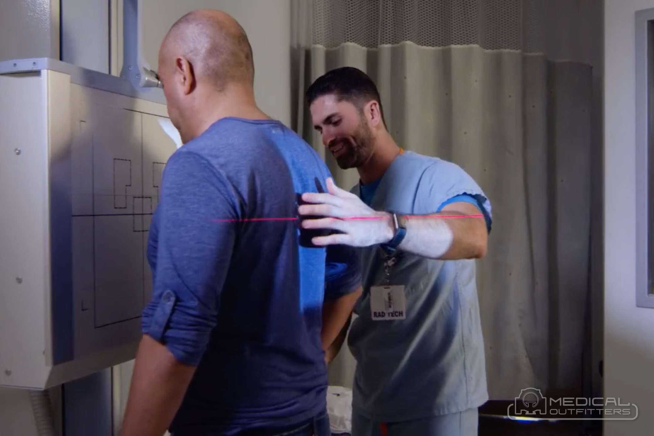
The sequence in the figure is the same in every class of system. The technologist selects the exam on the console, which loads the technique and, on an automated room, drives the tube and detector to position. The patient is positioned against the wall stand or on the table, the collimator is set to the body part, the exposure is made, and the panel converts the X-rays, reads out the pixel array and sends the raw data to the workstation. Processing for that body part is applied, the technologist checks positioning and the exposure index with the patient still in the room, repeats a view if needed, and sends the study. Medical Outfitters’ CR vs DR guide notes that departments which converted report fewer repeats because positioning errors are caught at the table rather than at the reader.
Flat-Panel Detectors: CsI vs GOS, Wired vs Wireless, Sizes and Weights
The flat panel detector is the component that makes a digital X-ray machine digital, and the Medical Outfitters X-ray machine cost guide calls it the largest single line in an X-ray quote after the generator. Most medical panels are indirect-conversion detectors: a scintillator layer converts X-rays into visible light, and a thin-film transistor array of photodiodes under it converts the light into charge, one pixel at a time. The scintillator is the first specification to read. Cesium iodide (CsI) grows as needle-like columns that guide the light to the pixel below, so it can be made thick enough to stop more X-rays without blurring the image. Gadolinium oxysulfide (GOS, also written Gd2O2S) is a granular phosphor screen that scatters its light, so it is kept thinner and converts less of the beam.
The difference is measured as detective quantum efficiency, the fraction of the information in the beam that reaches the image. A bench comparison of a CsI and a GOS flat panel from the University of Bologna, indexed in the EPA’s HERO database, measured peak DQE of 60, 60, 58 and 50 percent for CsI against 40, 44, 44 and 35 percent for GOS across four beam qualities, about 1.5 times higher for CsI, with modulation transfer at the Nyquist frequency of about 35 percent against 25 percent. Fujifilm quotes a DQE of 33 percent at 1 line pair per millimeter for its film-based GOS FDR D-EVO III panels, up from 31 percent for the glass-based generation, and describes its CsI panels as the higher DQE option for lower dose. In practice a CsI panel is specified where volume, dose or image quality matter, which is most rooms and carts, and a GOS panel where budget leads, such as a second panel for extremities or a low-volume room.
The second specification is wired or wireless. A tethered panel is fixed in the table or wall bucky and never runs out of battery; a wireless panel is the size of a film cassette, runs on a battery and moves between the table, the wall stand, a mobile cart and the patient’s bed. The third is size: 14 by 17 inches (35 by 43 cm) is the general-purpose standard, 17 by 17 inches (43 by 43 cm) covers chest and large fields without turning the panel, 10 by 12 inches (25 by 30 cm) suits extremities and pediatric work, and a 17 by 32 inch long panel takes a full spine or leg in one exposure. Fujifilm’s specification page for the FDR D-EVO III family, the panel Medical Outfitters supplies with its rooms and portables, gives the figures in the table.
| FDR D-EVO III model | Scintillator | Image area | Weight without battery | Pixels (0.15 mm pitch) |
|---|---|---|---|---|
| C35i (DR-ID 1811SE) | Cesium iodide | 14 x 17 in, 460 x 384 x 15 mm housing | About 2.2 kg (4.9 lb) | 2,836 x 2,336 |
| G35i (DR-ID 1831SE) | Gadolinium oxysulfide | 14 x 17 in, 460 x 384 x 15 mm housing | About 1.8 kg (4.0 lb) | 2,836 x 2,336 |
| C43i (DR-ID 1812SE) | Cesium iodide | 17 x 17 in, 460 x 460 x 15 mm housing | About 2.6 kg (5.7 lb) | 2,836 x 2,832 |
| G43i (DR-ID 1832SE) | Gadolinium oxysulfide | 17 x 17 in, 460 x 460 x 15 mm housing | About 2.1 kg (4.6 lb) | 2,836 x 2,832 |
| G80i (DR-ID 1836SE) | Gadolinium oxysulfide | 17 x 32 in, 850 x 460 mm housing | About 5.2 kg (11.5 lb) | 5,376 x 2,832 |
Source: Fujifilm FDR D-EVO III specification pages; the i-series panels use a flexible film-based transistor array rather than glass, connect over IEEE 802.11n and 802.11ac, and carry a battery pack of about 180 to 220 grams. Fujifilm’s irradiation side sampling (ISS) design places the transistor array on the side facing the tube so the light is captured before it spreads, which is where its DQE and sharpness figures come from.
Three more lines belong on the detector specification: the battery and how many exposures it gives per charge, the drop and liquid protection (wireless panels are dropped, and a dropped panel is the expensive failure, which is why Medical Outfitters’ CR vs DR guide recommends a drop warranty and a spare battery), and the exposure detection. A panel that detects the exposure itself can work with any generator, which is how a retrofit avoids rewiring an older room; a panel that is wired or wirelessly synchronized to the generator needs the interface. Panel lifetimes of seven to ten years and more are normal with careful handling, per the same guide, with the battery replaced every few years and the calibration checked at preventive maintenance.
The Generator, the Tube and the Stand That Carries It
The generator sets what the room can image and how fast. It is rated in kilowatts, and the Medical Outfitters X-ray machine cost guide draws the line at 32 kW for a clinic room against 65 to 82 kW for a hospital room that needs short exposures on large patients; Fujifilm’s FDR Smart Essential, which the guide prices in both configurations, is sold with a 32 kW generator on the floor-mounted stand and 65 to 82 kW on the ceiling-suspended one. A higher rating means a shorter exposure for the same technique, which matters for chest, abdomen and bariatric work where motion blurs the image. On a mobile cart the generator runs from the battery; Carestream’s DRX-Revolution carries a 32 kW generator, per Medical Outfitters’ portable X-ray price guide, which is why a full-size cart can cover an ICU while a handheld unit is limited to extremities, chest and bedside work.
The federal performance standard for radiographic equipment, 21 CFR 1020.31, is the specification sheet behind every generator and collimator sold in the United States, and its numbers explain what the physicist tests at acceptance. The exposure must be reproducible: the coefficient of variation of the air kerma over ten consecutive exposures may not exceed 0.05. It must be linear across the current settings, with the difference between two consecutive mAs settings held within 0.10 of their sum. The light field that the technologist uses to aim must match the X-ray field within 2 percent of the source-to-image distance, and the light localizer must deliver at least 160 lux at 100 cm. Positive beam limitation, where fitted, must block the exposure if the field exceeds the receptor by more than 3 percent of the SID in either dimension or 4 percent in total. Mobile and portable systems must prevent a source-to-skin distance shorter than 30 cm. Every system must show a visible signal when X-rays are produced and sound an audible one when the exposure ends. These are not features to compare between brands; they are the floor every cleared system meets.
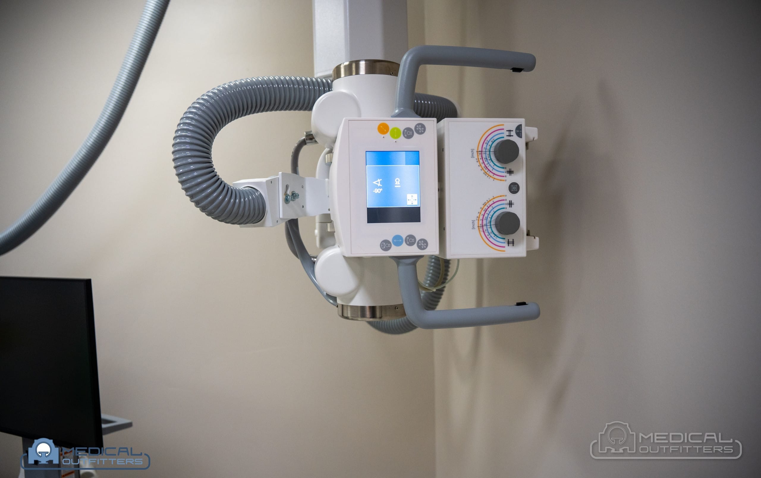
The stand is the part of a digital X-ray room that sets its tier, its room and most of the gap between a $60,000 room and a $275,000 one. The Medical Outfitters cost guide splits fixed rooms by it. A floor-mounted column runs on a floor track, needs no ceiling structure and anchors the entry tier; Fujifilm’s Essentia FS is the example the guide gives. A U-arm carries the tube and the detector on one stand that rotates around the patient, positioned by hand on an entry room such as the GE Definium 5000, per the GE Definium price guide. A straight arm links the tube and detector on a single arm; the Fujifilm Essentia SA installs under an 8 foot ceiling and weighs 551 pounds, per Medical Outfitters’ guide to X-ray room size, which is why it suits orthopedic and outpatient rooms with limited ceiling structure. A ceiling-suspended overhead tube crane runs on rails and reaches any position over the table and the wall stand, with automatic positioning on the premium rooms: Fujifilm’s FDR Visionary Suite moves the tube and the matching detector to the exam position and tracks manual movements of the table and wall stand, per Fujifilm’s US product page, and GE’s Definium 656 HD has five-axis motorization with auto-positioning, a 12 inch touchscreen at the tube and a 3D camera that checks positioning, per AuntMinnie’s report on its 2022 launch.
Types of X-Ray Machines: Room, Mobile Cart, Portable, Retrofit and R/F
The Medical Outfitters cost guide counts nine types of X-ray machine; for a buyer of general radiography they collapse into four purchases, plus the specialty rooms priced per project. The question arrives in many forms, types of X ray machines, DR X ray machine, digital X ray room, mobile X ray machine, portable digital X ray machine, and the four classes in the figure answer all of them.
The fixed DR room is the workhorse: table, wall stand, one or two panels, a generator in the equipment closet, and shielding designed to NCRP Report 147, which Medical Outfitters’ room guide names as the reference states adopt. It is sold in the three tiers above. New rooms from Medical Outfitters are Fujifilm: the Essentia FS and FDR Smart Essential at entry, the Essentia SA, Clinica X FS and ceiling-suspended Smart Essential in the middle, and the Clinica X OTC and FDR D-EVO Suite II at the top, with the FDR Visionary Suite as Fujifilm’s automated flagship. Refurbished rooms are sourced on request through MO Select: GE Definium 5000, 6000 and 8000, Discovery XR656 and 656 HD, Siemens Multix Fusion and Ysio Max, and Carestream DRX-Evolution, per the cost guide’s price list.
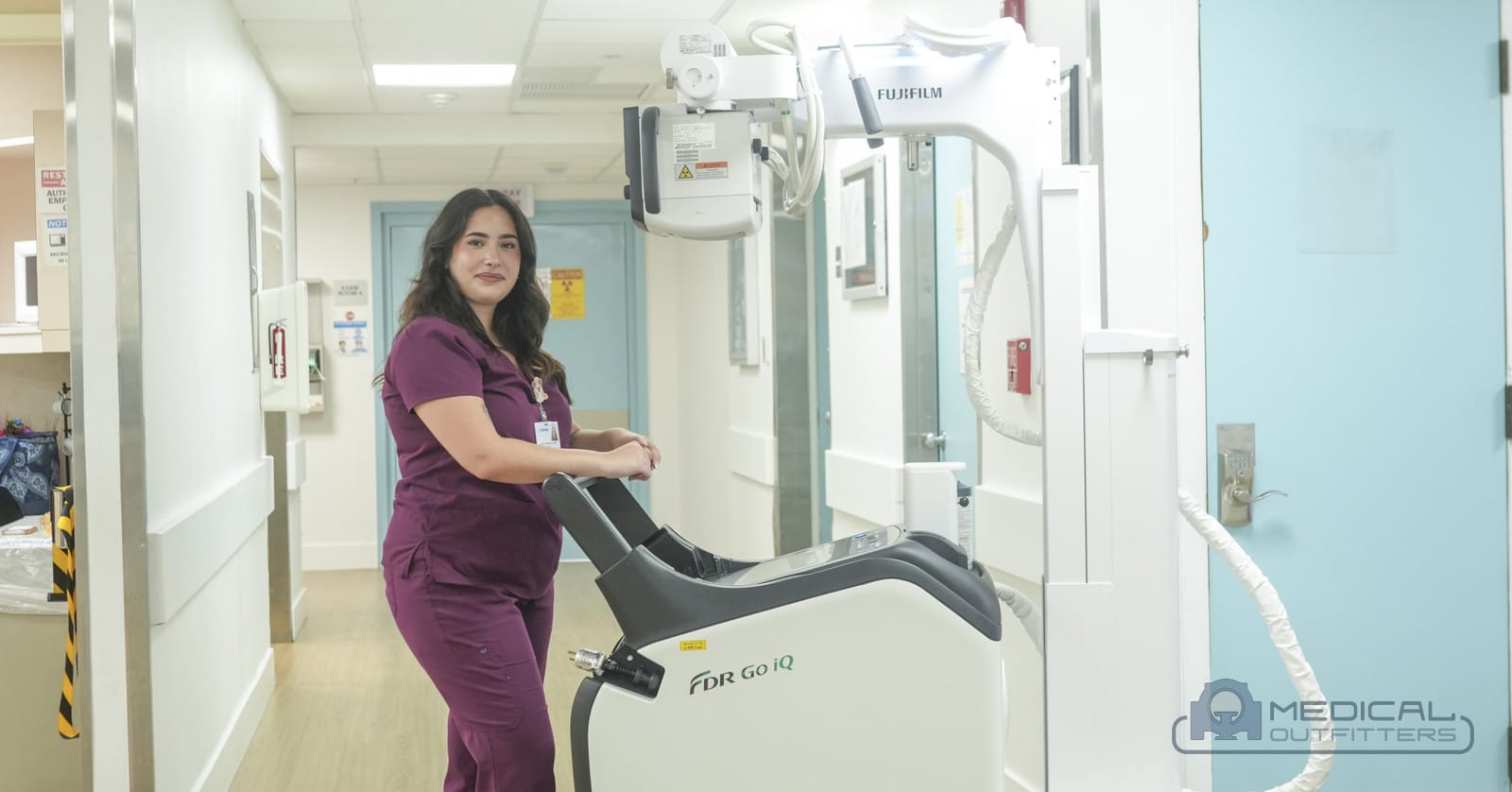
The full-size mobile cart brings the room to the ward, the ICU and the emergency department: a motorized, battery-driven cart with a hospital-grade generator, a wireless panel and PACS on board. Fujifilm’s FDR Go iQ, launched in the United States on July 30, 2025 per Fujifilm’s announcement, is 22.5 inches wide, weighs 970 pounds, carries a 19 inch display and a touchscreen at the tube head, and takes FDR D-EVO III panels in 14 by 17, 17 by 17, 10 by 12 and 17 by 32 inch sizes. Medical Outfitters demonstrated it at Larkin Community Hospital. The used market adds GE AMX and Optima, Siemens Mobilett, Carestream DRX-Revolution and Shimadzu MobileDaRt carts, priced by generation in the portable X-ray price guide.
The compact cart and the handheld unit cover the places a full-size cart cannot reach. Fujifilm’s FDR nano is a 90 kg cart with a wireless panel for corridors, elevators and nursing-home rooms, and the FDR AQRO is the compact cart in the photo below; the handheld FDR Xair weighs about 3.5 kg with its battery and takes up to 100 images on a charge with an FDR D-EVO II detector, per the portable guide, which is why home health, long-term care and field teams buy it. A handheld unit is not a room replacement: its output suits extremities, chest and bedside work, not thick-part imaging at volume.
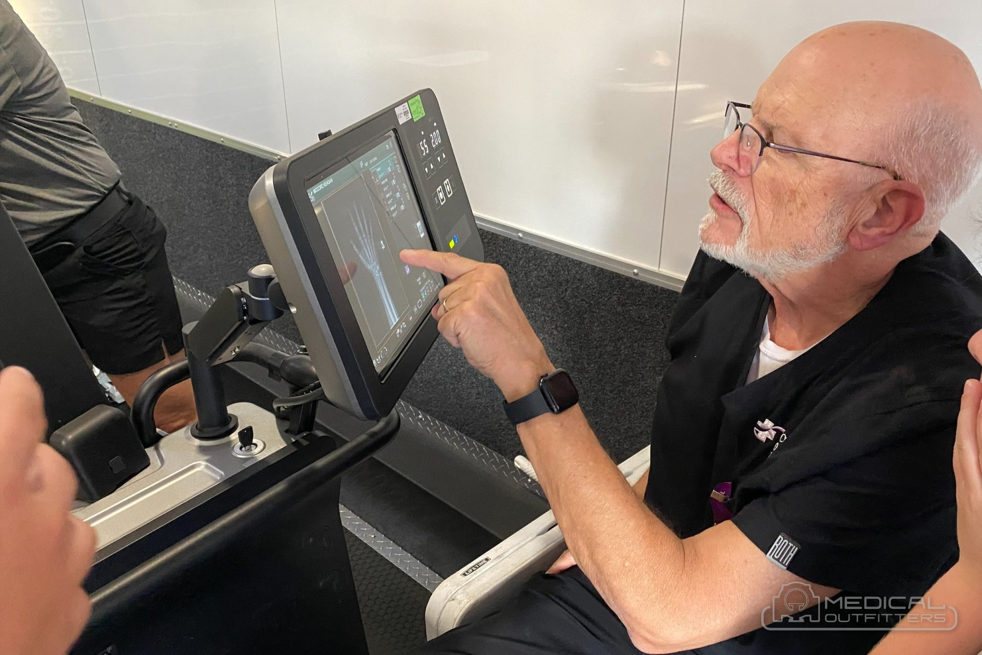
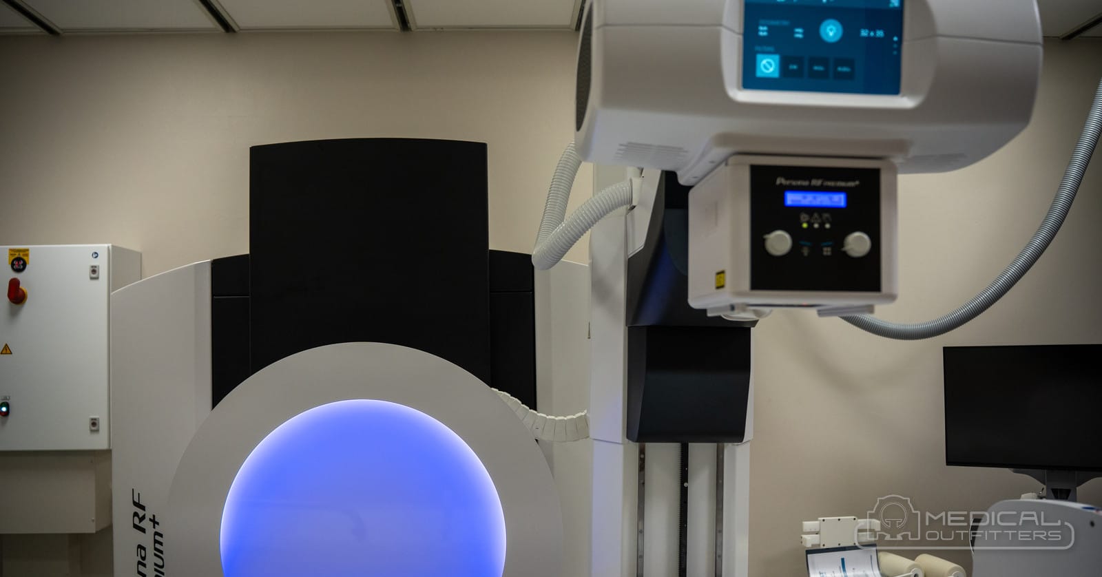
The DR retrofit is the fourth purchase and often the cheapest: a wireless panel, an acquisition workstation and either a generator interface or automatic exposure detection, added to a film or CR room whose generator and tube are sound. Medical Outfitters’ CR vs DR guide puts a complete retrofit at $25,000 to $60,000 delivered and installed, with the panel size, a second panel and the age of the generator setting the point in the range, and walks through the six steps from assessing the generator to retiring the cassettes. Radiography and fluoroscopy rooms such as the Fujifilm Persona RF PREMIUM+ and FDR Beyond add live imaging to a radiography room and are quoted per project, as are the FDR Cross hybrid C-arm and the VXR veterinary room; mammography, cone beam CT and dental units are X-ray machines too, but they are bought by other departments and priced in their own guides.
The Console: Image Processing, Exposure Index, Grid-Free Imaging and Stitching
The workstation is where a modern digital X-ray machine earns its keep after the exposure, and the software licenses on it move the quote, as the Medical Outfitters cost guide’s component table notes. The first job is body-part processing. Fujifilm’s FDX Console runs Dynamic Visualization II, which the company describes as recognizing bone, anatomy and orthopedic hardware and adjusting contrast and density by image thickness and structure, and GE’s Definium 656 HD runs Helix 2.2, which AuntMinnie reported as anatomy-specific enhancement across variations in dose, positioning, field of view and metal implants. The practical effect is an image that looks the same from one technologist to the next, which is the point: repeats fall when processing stops depending on technique.
The second job is keeping the dose honest. AAPM Task Group 116 (2009) standardized the exposure index, the number every DR console shows after an exposure, and the deviation index that compares it with the target the department sets per exam, building on the IEC 62494-1 definition; TG-232 (2018) revised the action levels to reflect clinical practice. Fujifilm’s FDX Console displays both and writes them into the DICOM header for dose monitoring, per Fujifilm’s product page. CR plates never look over-exposed because the reader rescales the image, which is why exposure crept upward in CR departments; a DR console with a deviation index shows the drift on every image.
The third job is the optional applications. Virtual grid software estimates and removes scatter from an image taken without a physical anti-scatter grid; Fujifilm reports a 50 percent dose reduction in its own CD-RAD phantom tests against an 8:1 grid, a vendor figure, and the benefit for bedside work is a lighter, faster exposure with no grid to align. Long-length stitching joins images for scoliosis and long-leg studies; Fujifilm’s Visionary Suite captures a full-length image in as little as 20 seconds, up to 63 inches upright and 47 inches on the table, per its US page, and GE’s Auto Image Paste and AutoSpine do the same on the 656 HD. Tomosynthesis reconstructs slices from a single sweep of the tube (Fujifilm’s Digital Tomosynthesis, GE’s VolumeRAD), and dual-energy subtraction takes two exposures at different energies to produce bone-only and soft-tissue-only views. Each is a license, each is a line on the quote, and a refurbished room may need a software reload or a license transfer before it has them, which the cost guide flags under the used-market checks.
The fourth job is connectivity: the modality worklist from the RIS, DICOM send to PACS, and the dose report. The Medical Outfitters cost guide lists the PACS integration among the items quoted per project, and the X-ray service page lists the detector calibration, the overhead crane and wall stand mechanics and the generator and tube as the service work a DR room needs over its life.
Radiation Dose From a Digital X-Ray, and Why Medicare Pays DR in Full
A digital X-ray is a low-dose exam, and the detector is the reason. RadiologyInfo, the patient site of the American College of Radiology and the Radiological Society of North America, lists the approximate effective doses in the table, with the equivalent period of natural background radiation; the FDA’s radiography page adds that the dose from radiography is generally less than from fluoroscopy and CT, that the benefit of a needed exam far exceeds the small cancer risk, and that exposure should be kept as low as possible with the fewest images needed.
| Exam | Approximate effective dose | Equivalent natural background |
|---|---|---|
| Chest X-ray | 0.1 mSv | 10 days |
| Lumbar spine X-ray | 1.4 mSv | 6 months |
| Extremity (hand, foot) X-ray | Less than 0.001 mSv | Less than 3 hours |
| Dental X-ray | 0.005 mSv | 1 day |
Source: RadiologyInfo radiation dose chart; typical values for an average-sized adult, varying with body size and imaging practice. Medical Outfitters’ CR vs DR guide cites published chest radiography comparisons that put the patient dose on an amorphous-silicon DR system at roughly 1.7 times lower than on CR and 2.7 times lower than film, which is the DQE difference described above turned into dose.
Payment follows the technology. Section 502 of the Consolidated Appropriations Act of 2016 cut the technical component of the Medicare payment for X-rays taken on film by 20 percent from January 1, 2017, reported with modifier FX, and for X-rays taken with computed radiography by 7 percent from 2018 through 2022 and 10 percent from January 1, 2023 onward, reported with modifier FY, per CMS transmittal R3583 and the implementing guidance. Digital radiography carries no modifier and no reduction. For a room that bills a few thousand Medicare X-rays a year, the CR reduction is a recurring cost that a DR purchase or retrofit removes in its first year, before any gain from throughput or repeat rate is counted.
Digital X Ray Machine Cost: What Each Class Costs in 2026
The class decides the band and the detector, generator and stand decide the point inside it. The figures below are Medical Outfitters all-in ranges from the X-ray machine cost guide, for a system delivered, installed and warrantied, with applications training; equipment-only prices published elsewhere stop before delivery, rigging, installation and current tariffs, so they read lower.
| Class | Refurbished, all-in | New, all-in | Who buys it |
|---|---|---|---|
| Portable or handheld DR | $12,000 to $35,000 | $30,000 to $65,000 | Home health, nursing facilities, bedside and field work |
| Mobile X-ray cart (full digital) | $40,000 to $90,000 | $90,000 to $185,000 | Hospital wards, ICU and emergency |
| Fixed DR room, entry tier (floor column or U-arm) | $60,000 to $95,000 | $150,000 to $185,000 | Clinics, urgent care, orthopedic and chiropractic practices |
| Fixed DR room, mid tier (straight arm or crane-ready floor stand) | $70,000 to $115,000 | $160,000 to $220,000 | Imaging centers and multi-doctor groups |
| Fixed DR room, premium tier (ceiling-suspended, automated) | $95,000 to $135,000 | $200,000 to $275,000 | Hospitals and high-volume departments |
| DR retrofit of a working room | $25,000 to $60,000, panel, workstation, integration, installation and training | Any film or CR room with a sound generator | |
Source: Medical Outfitters X-ray machine cost guide and CR vs DR guide, reviewed September and October 2026. New rooms are the Fujifilm systems Medical Outfitters sells as an authorized dealer; refurbished rooms are GE, Siemens and other makes sourced and rebuilt on request through MO Select. The current GE flagship sits above the standard band: the Definium price guide lists a refurbished or pre-owned Definium 656 HD at $135,000 to $225,000 and a new one through GE at an estimated $425,000 to $850,000, a comparison figure and not a Medical Outfitters offer.
What sits inside the number matters as much as the number. The cost guide’s component table lists what a Medical Outfitters price includes: the generator, the tube and its stand, the table and wall stand where specified, the detector and workstation, the software as listed, delivery, rigging, installation, calibration, applications training and the written warranty. Quoted per project, because they depend on the building, are the lead shielding designed to NCRP Report 147, the control booth and leaded window, the electrical feed, the physicist’s acceptance survey, the state registration, the PACS integration and the first service contract. Two quotes that differ by tens of thousands of dollars usually differ in that list rather than in the machine.
How to Choose a Digital Radiography System
Medical Outfitters’ X-ray category page settles most purchases with four questions before a model is named: where the system will be used, how many exams a day, DR or CR, and new, refurbished or used. The answers map onto the parts in this guide.
Where decides the class. A dedicated room with a table and wall stand is a fixed DR system and brings shielding and a control booth with it; imaging where the patient is means a mobile cart or a handheld unit and no site work. A hospital usually buys both, the cost guide notes: a premium room with two or three carts for the wards.
Volume decides the generator and the stand. The cost guide’s tiers put a floor column with one panel at about 7 to 15 patients a day, a straight arm or crane-ready floor stand at 15 to 25, and an overhead crane with automatic positioning and two panels at 30 or more. A room that positions itself and never waits for a panel to be moved turns over faster, which is what the premium tier buys.
The detector decides image quality and dose. Specify the scintillator, the size, wireless or tethered, one panel or two, and the drop and battery warranty, in that order. A second fixed panel in the wall stand removes panel swapping on a busy room; a single wireless panel shared between table, wall stand and a cart is the economical choice for a clinic.
Condition decides the warranty and the software. New Fujifilm systems come with the manufacturer’s warranty and current software; a refurbished room from MO Select is inspected, tested and sold with a written warranty, and the cost guide’s used-market section names what sets a used price before the model does: the detector, the generator and tube condition, and whether the manufacturer still supports the software, with a software reload or license transfer sometimes needed. Medical Outfitters’ used imaging equipment inspection checklist covers the same ground for a system bought elsewhere. For a room that already has a sound generator, the retrofit path is the cheapest way to a DR department, and the CR vs DR guide gives the six steps.
Buying a Digital X-Ray Machine With Medical Outfitters
Medical Outfitters sells new Fujifilm digital radiography rooms from the floor-mounted FDR Smart Essential to the ceiling-suspended FDR D-EVO Suite II, the FDR portables and compact carts, and the FDR D-EVO III wireless detector on its own for a retrofit, and it sources refurbished GE and Siemens rooms through MO Select, priced model by model in the X-ray machine cost guide, the portable X-ray machine price guide and the GE Definium price guide. The X-ray machines for sale page lists the current systems, and the X-ray service team maintains rooms and carts we sold and rooms bought elsewhere. Every quote covers delivery, rigging, installation, calibration, applications training and a written warranty, across the United States, Puerto Rico and the Caribbean. For a price range on a room, a cart or a retrofit, send the exam mix, the room drawing and, for a retrofit, the generator make, model and age to Medical Outfitters.
- US FDA, Radiography: the definition of radiography, its uses, risks and the applicable performance standards, 21 CFR 1020.30 and 1020.31. fda.gov
- 21 CFR 1020.31, Radiographic equipment (Cornell Legal Information Institute): reproducibility, linearity, field alignment, positive beam limitation, minimum source-to-skin distance and beam-on indication limits. law.cornell.edu
- RadiologyInfo, Radiation Dose: approximate effective doses for chest, lumbar spine, extremity and dental X-rays. radiologyinfo.org
- Fujifilm, FDR D-EVO III specifications (C35i, G35i, C43i, G43i, G80i): scintillator, size, weight, pixel pitch and wireless standard; FDR D-EVO III G35/G43 page: DQE 33 percent and the film-based transistor array. fujifilm.com
- Fujifilm Healthcare Americas, FDR Visionary Suite and FDX Console: auto-positioning, tube tracking, long-length stitching, tomosynthesis, dual energy, Dynamic Visualization II, Virtual Grid and the exposure and deviation index. healthcaresolutions-us.fujifilm.com
- Fujifilm Healthcare Americas, FDR Go iQ launch announcement, July 30, 2025: width, weight, display and detector sizes. businesswire.com
- AuntMinnie, GE releases new Definium 656 HD flagship fixed X-ray system, August 1, 2022: five-axis auto-positioning, 100 micron FlashPad HD detectors, Helix 2.2, VolumeRAD. auntminnie.com
- University of Bologna bench comparison of CsI and GOS flat-panel detectors, indexed in the EPA HERO database: peak DQE and MTF figures. hero.epa.gov
- AAPM Task Group 116 (2009) and Task Group 232 (2018): the exposure index and deviation index for digital radiography. aapm.org
- CMS transmittal R3583CP and Section 502 of the Consolidated Appropriations Act of 2016: the film and computed radiography payment reductions and modifiers FX and FY. cms.gov
- X-ray machine cost and price guide (2026)
- Portable X-ray machine price guide (2026)
- GE Definium X-ray price guide (2026)
- X-ray machines for sale: Fujifilm FDR, new and refurbished
- X-ray machines: rooms, portables and R/F systems
- Digital detectors and flat panels
- Fujifilm FDR D-EVO III wireless flat-panel detector
- Fujifilm FDR Smart Essential digital radiography room
- Fujifilm FDR D-EVO Suite II DR room
- X-ray machine service, repair and DR upgrades
- CR vs DR X-ray: how they differ and what a retrofit costs
- What size room an X-ray machine needs
- The Fujifilm FDR Go iQ mobile X-ray demonstration at Larkin
- Radiology equipment cost in 2026, every modality
- Ask for a digital X-ray price range
Digital X-Ray Machines: Common Questions
What is a digital X-ray machine?
A digital X-ray machine is a radiography system that records the image on an electronic flat-panel detector instead of film or a cassette, so the image appears on the console two to five seconds after the exposure. The FDA defines radiography as creating and recording an X-ray pattern as a still image and regulates the equipment under 21 CFR 1020.30 and 1020.31. The machine is a generator, a tube on a stand, a detector and a workstation.
What is the difference between a digital X-ray machine and a conventional one?
A conventional machine records the image on film that is developed, or on a computed radiography cassette that is scanned by a reader; a digital radiography machine reads the image straight from a flat panel. The generator and tube are often the same, which is why a working film or CR room can be converted by adding a panel and a workstation, a retrofit the Medical Outfitters CR vs DR guide prices at $25,000 to $60,000 installed.
How much does a digital X-ray machine cost?
The Medical Outfitters X-ray machine cost guide gives all-in ranges for 2026: $12,000 to $35,000 refurbished and $30,000 to $65,000 new for a portable or handheld unit, $40,000 to $90,000 refurbished and $90,000 to $185,000 new for a full-size mobile cart, and $60,000 to $135,000 refurbished and $150,000 to $275,000 new for a fixed DR room, delivered, installed and warrantied. Shielding and the control booth are quoted per project.
What is the difference between CsI and GOS detectors?
Both are scintillators that turn X-rays into light for the pixel array beneath. Cesium iodide grows as columns that guide the light, so it can be thick and still sharp; gadolinium oxysulfide is a granular screen that scatters light and is kept thin. A University of Bologna bench comparison measured about 1.5 times the detective quantum efficiency for CsI, which means lower dose or a cleaner image. CsI is the choice for most rooms and carts, GOS for budget and extremity panels.
Are wireless X-ray detectors better than wired ones?
They are more flexible, not better at imaging. A wireless panel is cassette-sized, battery powered and moves between the table, the wall stand, a cart and the bedside, so one panel can serve a clinic room and a portable. A tethered panel stays in its bucky and never runs out of charge. Fujifilm's wireless FDR D-EVO III C35i weighs about 2.2 kg without its battery, per Fujifilm's specification page; the expensive failure is a drop, so a drop warranty matters.
How much radiation does a digital X-ray give?
Less than fluoroscopy or CT, per the FDA. RadiologyInfo lists a chest X-ray at about 0.1 mSv, the equivalent of 10 days of natural background radiation, a lumbar spine series at 1.4 mSv and a hand or foot X-ray at less than 0.001 mSv. Digital detectors need less exposure than film or CR for the same image because they convert more of the beam, and the exposure index on every image shows the technologist when technique drifts.
Does Medicare pay less for digital X-rays?
No, the reductions go the other way. Section 502 of the Consolidated Appropriations Act of 2016 cut the technical component for X-rays taken on film by 20 percent from 2017, reported with modifier FX, and for computed radiography by 7 percent from 2018 and 10 percent from 2023 onward, reported with modifier FY. Digital radiography is paid in full with no modifier, which is one of the arithmetic lines behind every DR retrofit.
What size generator does a digital X-ray room need?
The Medical Outfitters X-ray machine cost guide draws the line at 32 kW for a clinic room and 65 to 82 kW for a hospital room that needs short exposures on large patients. Fujifilm's FDR Smart Essential is sold with a 32 kW generator on the floor-mounted stand and 65 to 82 kW on the ceiling-suspended one. A higher rating shortens the exposure for the same technique, which reduces motion blur on chest, abdomen and bariatric work.
Related Articles